Practical anatomy: a manual of dissections / by Christopher Heath.
- Christopher Heath
- Date:
- 1870
Licence: Public Domain Mark
Credit: Practical anatomy: a manual of dissections / by Christopher Heath. Source: Wellcome Collection.
Provider: This material has been provided by the Francis A. Countway Library of Medicine, through the Medical Heritage Library. The original may be consulted at the Francis A. Countway Library of Medicine, Harvard Medical School.
441/600 (page 431)
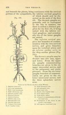
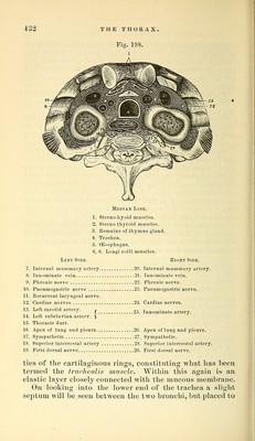
![Splanchnic Nerves (Fig. 194, 43, p. 420).—The great splanchnic nerve is derived from four ganglia (*7th, 8th, 9th, 10th) by separate fibres. The nerve runs inwards and thus enters the lower part of the mediastinum, and after pierc- ing the crus of the diaphragm joins the solar plexus in the abdomen. The lesser splanchnic nerve is derived from the 10th and llth ganglia, and also pierces the crus of the diaphragm to join the solar or renal plexus. The least [or renal'] splanchnic nerve is derived from the 12th ganglion, and goes to the renal plexus. It is seldom found, in which case the lesser nerve is connected with the ganglion. The Internal Intercostal Muscles (Fig. 195, p. 424) can be seen beneath the pleura without any further dissec- tion. Beginning at the sternum the muscles reach as far as the angles of the ribs, at which points the intercostal vessels and nerves h'ing against the external intei'costals are visible. The fibres of the internal intercostals take a direction contrary to that of the external intercostal mus- cles, i.e., the}^ run forwards and upwards [like the fibres of the internal oblique]. The relation of the parts passing through the superior aperture of the thorax can be now full}^ understood, and will be found in the following table and the accompanying diagram taken from nature (iFig. 198). The lungs which have been removed and laid aside are now to be dissected, and the structure of the trachea and lungs is to be exa- mined. The Trachea is about four inches and a half in length, and is convex in front but flattened posteriorly, being com- posed of a series of cartilages, the extremities of which are connected behind by fibrous and muscular tissue. There are from sixteen to twenty cartilages, each measuring about two lines in depth but decreasing in depth from above downwards. The last cartilage is peculiar, in being cut obliquely on each side so as to be adapted to the com- mencement of the bronchi. The cartilages are connected together by fibrous tissue, and the first is similarly con- nected to the cricoid cartilage. On dissecting away the fibrous tissue at the back of the trachea, together with numerous mucous glands, involun- tary muscular fibres will be seen connecting the extremi-](https://iiif.wellcomecollection.org/image/b21057679_0441.jp2/full/800%2C/0/default.jpg)