Practical anatomy: a manual of dissections / by Christopher Heath.
- Christopher Heath
- Date:
- 1870
Licence: Public Domain Mark
Credit: Practical anatomy: a manual of dissections / by Christopher Heath. Source: Wellcome Collection.
Provider: This material has been provided by the Francis A. Countway Library of Medicine, through the Medical Heritage Library. The original may be consulted at the Francis A. Countway Library of Medicine, Harvard Medical School.
444/600 (page 434)
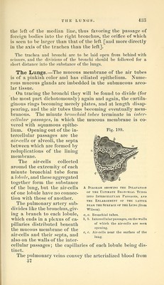
![the lobules, and correspond to the branches of the arteries. They have no valves. The bronchial arteries may be traced upon the bronchial tubes for some distance. They supply the substance of the lung, and their blood is returned partly by the bronchial veins and partly by the pulmonary veins. (Waters.) Prevertebral Region. The carotid arteries with the jugular veins, and tlie pneumogastric and sympathetic nerves, are to be divided at the level of the top of the sternum, and the trachea with the oesophagus is to be severed a little lower down. The neck is then to be bent forcibly backward so as to make the cut surface of the sliull rest upon the table, and the cesophagus and trachea with the vessels and nerves being drawn forcibly upward, the cellular tissue between the pharynx and the front of the vertebral column is to be cautiously dissected through until the under surface of the base of the skull is exposed. The saw is now to be applied close behind the mastoid process and an oblique cut made, which is to be carried through the whole thickness of the temporal bone into the jugular foramen, and prolonged through the remaining portion of the parietal bone to the cut which was made in removing the brain. A similar cut having been made on the opposite side, a broad chisel is to be applied to the basilar process of the occi- pital bone where it is exposed behind the pharynx, and it is to be divided. The chisel being again applied on each side of the middle line will unite this cut with those made by the saw, and the prepara- tion will then be divided into two parts; the anterior part of the skull with the pharynx and deep vessels and nerves is to be wrapped up for subsequent examination [p. 436], and the muscles attached to the vertebral column with the posterior part of the skull are now to be examined. The Scaleni muscles have been seen already in part, but can now be fully dissected. The Scalenus Anticus (Fig. 200, 2) arises from the tubercle on the inner border and upper surface of the first rib (scalene tubercle), and ascends to be inserted into the anterior tubercles of the transverse processes of the 3d, 4th, 5th, and Gth cervical vertebrae. The phi'enic nerve will pro- bablj'' still be found on the anterior surface of the muscle, and behind it the brachial nerves emerge and the subcla- vian artery passes. The Scalenus Medius (Fig. 200, 7) lies behind the brachial nerves, arising from the rough marking upon the upper surface of the first rib behind the groove for the sub- clavian artery. It ascends to be inserted into the poste-](https://iiif.wellcomecollection.org/image/b21057679_0444.jp2/full/800%2C/0/default.jpg)