Practical anatomy: a manual of dissections / by Christopher Heath.
- Christopher Heath
- Date:
- 1870
Licence: Public Domain Mark
Credit: Practical anatomy: a manual of dissections / by Christopher Heath. Source: Wellcome Collection.
Provider: This material has been provided by the Francis A. Countway Library of Medicine, through the Medical Heritage Library. The original may be consulted at the Francis A. Countway Library of Medicine, Harvard Medical School.
447/600 (page 437)
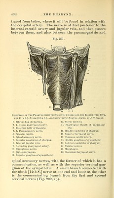
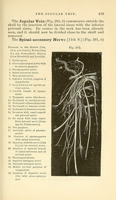
![l\y the palato-glossns muscle. The posterior pillar passes oblique]}- backwards and is lost in the phar3'nx, being formed by the palato-phar^nigeus muscle. The tonsil is generally much shrunken in a subject which has arrived at this stage of dissection. The pharynx and upper part of the oesophagus are to be carefully distended with cotton-wool or tow, and the preparation being placed with the face downwards, is to be secured over a small block with hooks, one set of which should draw the cesophagus down and keep the pharynx tense. The vessels and nerves at the back of the pharynx are to be examined before the muscular bag itself is dissected. The vessels and nerves now to be examined have all been seen in part in previous dissections, and then from either the front or the side. They are now all seen from behind, and this must be borne in mind thoroughly, or will lead to misconception of the description. The section of the base of the skull is seldom precisely similar on the two sides and it will generally be found advisable therefore to trace the parts first brought into view on one side, and the carotid artery, etc., on the other, as in the illustration (Fig. 201, p. 438). The Sympathetic Nerve (Fig. 201, 12, 18) with its superior and middle cervical ganglia is at once exposed, and some of its branches ma}- be very conveniently traced. The superior cervical ganglion (i 2) is fusiform and nearly an inch in length. It lies behind the internal carotid artery, and has small branches of communication with the follow- ing cranial nerves—the glosso-pharyngeal, the pneumogas- tric, and the hypoglossal [Fig. 163, p. 300]. The branches of communication with the cervical nerves have been already seen (p. 359). The branches of distribution are, (1) the nervi molles distributed upon the external carotid artery and its branches; (2) the pharyngeal branch which can now be traced to the pharynx, where it enters in the forma- tion of the pharyngeal plexus; (3) the laryngeal branch to the superior lar3^ngeal nerve; (4) the superior cardiac nerve which has been already seen. The Middle cervical ganglion (18) is of small size, and gives off (1) thj-roid branches upon the inferior thyroid artery and (2) the middle cardiac nerve. The Ninth [12th S.) or Hypoglossal Nerve (Fig. 201, 10] is necessarily cut off at the anterior condyloid foramen in making the dissection, and should therefore be 37*](https://iiif.wellcomecollection.org/image/b21057679_0447.jp2/full/800%2C/0/default.jpg)