Practical anatomy: a manual of dissections / by Christopher Heath.
- Christopher Heath
- Date:
- 1870
Licence: Public Domain Mark
Credit: Practical anatomy: a manual of dissections / by Christopher Heath. Source: Wellcome Collection.
Provider: This material has been provided by the Francis A. Countway Library of Medicine, through the Medical Heritage Library. The original may be consulted at the Francis A. Countway Library of Medicine, Harvard Medical School.
448/600 (page 438)
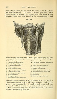
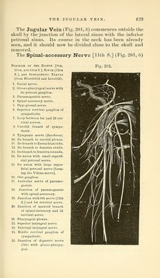
![traced from below, where it will be found in relation with the occipital artery. The nerve is at first posterior to the internal carotid artery and jugular vein, and then passes between them, and also between the pneumogastric and Fig. 201. Dis A 1. 2, 3. 4, 5. 6. 7. SECTTON OP THR PHARYNX WITH THi: CAROTID VESSELS AND THE EIGHTH [9TH, IOTF, ND llTH S.], Ninth [12ih S,], and Sympathetic Nerves (drawn by J. T. Gray). Fibrous bag of pharynx. 2. Glosso-pharyngeal nerve. Posterior belly of digastric. i. Pneumogastric nerve. Splenius capitis. Spinal-accessory nerve. Superior constrictor of pharynx. Internal jugular vein. Ascending pharyngeal artery. Hypoglossal nerve. Stylo-pharyngeus. Superior ganglion of sympathetic. 13. Sterno-mastoid. 14. Pharyngeal branch of pneumogas- tric. 15. Middle constrictor of pharynx. 16. Superior laryngeal nerve. 17. Common carotid artery. IS. Middle ganglion of sympathetic. 19. Inferior constrictor of pharynx. 20 Cardiac nerves. 21. (Esophagus. 22. Recurrent laryngeal nerve. spinal-accessory nerves, with the former of which it has a communication, as well as with the superior cervical gan- glion of the sympathetic. A small branch connected with the ninth [12th S.] nerve at one end and loose at the other is the communicating branch from the first and second cervical nerves (Fig. 202, 19).](https://iiif.wellcomecollection.org/image/b21057679_0448.jp2/full/800%2C/0/default.jpg)