Practical anatomy: a manual of dissections / by Christopher Heath.
- Christopher Heath
- Date:
- 1870
Licence: Public Domain Mark
Credit: Practical anatomy: a manual of dissections / by Christopher Heath. Source: Wellcome Collection.
Provider: This material has been provided by the Francis A. Countway Library of Medicine, through the Medical Heritage Library. The original may be consulted at the Francis A. Countway Library of Medicine, Harvard Medical School.
477/600 (page 467)
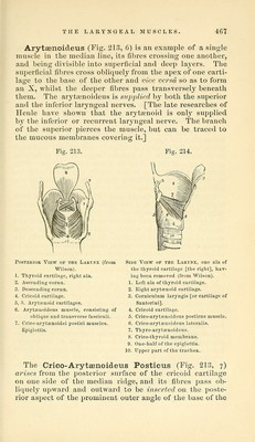
![THE LARYNGEAL MUSCLES. 46Y Arytaenoideus (Fig. 213, 6) is an example of a single muscle in the median line, its fibres crossing one another, and being divisible into superficial and deep layers. The superficial fibres cross obliquely from the apex of one carti- lage to the base of the other and vice versa so as to form an X, whilst the deeper fibres pass transversely beneath them. The arj-^ti^^noideus is siqDplied b_y both the superior and the inferior laryngeal nerves. [The late researches of Henle have shown that the arytenoid is onlj' supplied b} the inferior or recurrent larjaigeal nerve. The branch of the superior pierces the muscle, but can be traced to the mucous membranes covering it.] Fig. 213. Fig. 214. Posterior View of the Lartnx (from Wilson). 1. Thyroid cartilage, right ala. 2. Ascending cornii. 3. Descending cornu. 4. Cricoid cartilage. 5. ri. Arytenoid cartilages. 6. Arytcenoideus muscle, consisting of oblique and transverse fasciculi. 7. Crico-arytffinoidei postici muscles. •Epiglottis. Side View of the Larynx, one ala of the thyroid cartilage [the right], hav- ing heen removed (from Wilson). 1. Left ala of thyroid cartilage. 2. Eight arytenoid cartilage. 3. Corniculum laryngis [or cartilage of Santorini]. 4. Cricoid cartilage. 5. Crico-aryta3noideus posticus muscle. 6. Crico-arytsenoideus lateralis. 7. Thyro-arytcenoideus. 8. Crico-thyroid membrane. 9. One-half of the epiglottis. 10. Upper part of the trachea. The Crico-Arytsenoideus Posticus (Fig. 213, 7) arises from the posterior surface of the cricoid cartilage on one side of the median ridge, and its fibres pass ob- liquely upward and outward to be inserted on the poste- rior aspect of the prominent outer angle of the base of the](https://iiif.wellcomecollection.org/image/b21057679_0477.jp2/full/800%2C/0/default.jpg)