Practical anatomy: a manual of dissections / by Christopher Heath.
- Christopher Heath
- Date:
- 1870
Licence: Public Domain Mark
Credit: Practical anatomy: a manual of dissections / by Christopher Heath. Source: Wellcome Collection.
Provider: This material has been provided by the Francis A. Countway Library of Medicine, through the Medical Heritage Library. The original may be consulted at the Francis A. Countway Library of Medicine, Harvard Medical School.
483/600 (page 473)
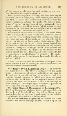
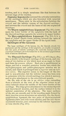
![The Cricoid Cartilage (Fig. 214, 4) (xpi»:oj fISoj, like a ring) is a ring of cartilage, shallow in front (where it has been seen to be connected with the thyroid cartilage by a membrane, and has the erico-thja-oideus attached to its surface), but deep behind, where it fills up a part of the space left between the posterior borders of the thyroid. The upper border of the deep portion presents two oval articular surfaces for the arytaenoid cartilages, external to which are the origins of the crico-arj'tsenoidei laterales. On each side and near the lower border of the cartilage are two small facets for articulation with the inferior cornua of the thjn'oid cartilage. The posterior surface is divided in the middle line by a vertical ridge to which some of the fibres of the oesophagus are attached, the concave surface on each side giving origin to the crico-arytsenoidei postici muscles. The Arytaenoid Cartilages (Fig. 215, d) (apuT-ai-Va, a pitcher^) are two in number, and are of a pyramidal shape. The base of each cartilage is triangular and articulates with the upper border of the cricoid cartilage; its anterior angle gives attachment to the true vocal chord, and its external angle to the crico-arytaenoideus posticus and crico-arj'tffinoideus lateralis. The apex is curved backward and inward, and is sur- mounted by the corniculum larj-ngis. The posterior sur- face of the cartilage is concave and gives attachment to the aryttfinoideus muscle; the anterior surface presents a small tubercle for the attachment of the false vocal chord, and also gives attachment to the thyro-ary ta^noideus muscle; the internal surface is smooth and covered with mucous membrane. The Cornicula Laryngis or Cartilages of Santorini (Fig. 215, y) are two small conical cartilages connected with the apices of the arytenoid cartilages and with the arytffino-epiglottidean folds. They are composed of yellow fibro-cartilage. The Cuneiform Cartilages [cartilages of Wrisberg] (Fig. 215, g) are two small bodies found in the arytffino- ' This derivation lias reference to the appearance of both cartilages taken together and covered by mucons membrane. In animals, which were the principal subjects of dissection among the ancients, the opening of the larynx, with the arytenoid cartilages, bears a curious resemblance to the mouth of a pitcher with a large spout (Wilson). 40*](https://iiif.wellcomecollection.org/image/b21057679_0483.jp2/full/800%2C/0/default.jpg)