Practical anatomy: a manual of dissections / by Christopher Heath.
- Christopher Heath
- Date:
- 1870
Licence: Public Domain Mark
Credit: Practical anatomy: a manual of dissections / by Christopher Heath. Source: Wellcome Collection.
Provider: This material has been provided by the Francis A. Countway Library of Medicine, through the Medical Heritage Library. The original may be consulted at the Francis A. Countway Library of Medicine, Harvard Medical School.
504/600 (page 494)
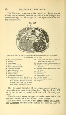
![The External brandies of the lower six dorsal and of all the lumbar nerves, become cutaneous at an oblique line corresponding to the margin of the aponeurosis of the latissimus dorsi. Section of Neck at the Fourth Cervical [Compare Fig. 155 1. Anterior jugular vein. 12. 2. Trachea. 3. CEsopliagus. - 13. 4. Steriio-hyoid muscle. 14 5. Sterno-thyroid muscle. 6. Platysma myoides. 15. 7. External jugular vein. 8. Sterno-mastoid. 16. 9. Thyroid gland. 17 10. Common carotid artery. 18. 11. Internal jugular vein. The de.scen- 19. dens noni nerve is seen in front of 20, these vessels, the pneumogastric 21 between them, and the sympa- thetic behind [and within] them. Vertebra (altered from Bdraud). , p. 338.] Longns colli, with rectus capitis an- ticus major to outer side. Scalenus anticuswith phrenic nerve. . Vertebral vessels in transverse process. Scalenus medius with brachial cords in front. Splenius colli. . Complexus. Semi-spinalis colli. Splenius capitis. . Levator anguli scapulse. Trapezius. The External branches of the upper sacral nerves be- come cutaneous near the median line. The fourth and fifth sacral nerves are very small and join with the coccygeal nerve. The Coccygeal nerve emerges from the lower end of the vertebral canal and is distributed to the coccyx. The Posterior Divisions of the Intercostal and Lum- bar Arteries divide like the nerves into internal and ex-](https://iiif.wellcomecollection.org/image/b21057679_0504.jp2/full/800%2C/0/default.jpg)