Practical anatomy: a manual of dissections / by Christopher Heath.
- Christopher Heath
- Date:
- 1870
Licence: Public Domain Mark
Credit: Practical anatomy: a manual of dissections / by Christopher Heath. Source: Wellcome Collection.
Provider: This material has been provided by the Francis A. Countway Library of Medicine, through the Medical Heritage Library. The original may be consulted at the Francis A. Countway Library of Medicine, Harvard Medical School.
505/600 (page 495)
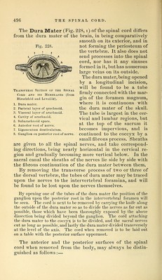
![tevnal bvaiiclies, which accompany the nerves to the mus- cles of the back. The Sixth Layer of Muscles (Fig. 225, p. 489) con- sists of the Interspinales, Intertransversales, Multifidus Spin;^, and Levatores Costarum, which will not repay the trouble of dissection. The position of the Interspinales and Intertransversales (iS) is sufficiently indicated by their names. The Multifidus Spinse (i6) fills up the vertebral groove beneath the erector spinse, arising from the articular pro- cesses of the cervical and lumbar vertebrae and from the transverse processes of the dorsal vertebrae and sacrum. The muscle is inserted into the spinous processes of all the vertebrae except the atlas. The Levatores Costarum are twelve fan-shaped muscles passing between the dorsal transverse processjes and the upper borders of the ribs. The Spinal Cord and Membranes. To open the spinal canal the remains of the muscles of the back should be cleared away as far as possible, when some part of the plexus of dor si-spinal veins may be seen upon the vertebrae. A block then being placed beneath the thorax so as to make the dorsal region prominent, a cut is to be made with the saw on each side of the middle line, so as to divide the laminae of the vertebrae as far out as possible. Two or three of the arches being now removed with the chisel, the point of a spine chisel or rachet is to be introduced into the canal and the rachet carefully hammered through the arches of the vertebraj for the whole length of the spine [the saw can also be used instead of the rachet] except the upper two cervical verte- brae. The operation being repeated on the opposite side, the arches can be removed with the bone-forceps, and the canal will be thoroughly opened. On the inner surface of the arches will be seen the liga- menta subjiava which are described with the other vertebral liga- ments (p. 476). Upon opening the Spinal Canal some loose tissue and fat will be seen, together with the meningo-racliidian veins, which are occasionally full of blood. These extend for the whole length of the spinal cord under the name of posterior longitudinal spinal veins, and communicate both with the veins outside the vertebrae and with the anterior longitudinal spinal veins at the backs of the bodies of the vertebrae. By removing the fat and veins the dura mater will be exposed.](https://iiif.wellcomecollection.org/image/b21057679_0505.jp2/full/800%2C/0/default.jpg)