Practical anatomy: a manual of dissections / by Christopher Heath.
- Christopher Heath
- Date:
- 1870
Licence: Public Domain Mark
Credit: Practical anatomy: a manual of dissections / by Christopher Heath. Source: Wellcome Collection.
Provider: This material has been provided by the Francis A. Countway Library of Medicine, through the Medical Heritage Library. The original may be consulted at the Francis A. Countway Library of Medicine, Harvard Medical School.
535/600 (page 525)
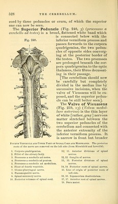
![aquecliK't of S^'lvius, which passes beneath the posterior commissure, the pineal gland and the corpora quadrigemina (Fig. 242, 16). In the tcetus the [third] ventricle commu- nicated in addition with the fifth ventricle and [also] with the infundibulum [by the iter ad infundibulum, the open- ing of which is just under the anterior commissure]. The Thalamus Opticus (Fig. 239, 6) is now fully ex- posed, and will be seen to be a lai'ge white body placed posteriorly to the corpus striatum and at the side of the third ventricle. [It is the great ganglion of sensation']. It has been seen to form part of the floor of the lateral ven- tricle by its upper surface, on which is a slight prominence called the anterior tubercle. Along the inner margin is a narrow white band, one of the peduncles of the pineal bod}^, and b}^ its inner surface which bounds the third ven- tricle, it gives attachment to the middle and posterior commissures of the third ventricle, the posterior piercing its substance. The thalamus opticus forms the roof of the descending cornu of the lateral ventricle, and by drawing it upward on the side upon which the cornu has been opened, two projections on its under surface may be seen. These are the Corpora Geniculata (externum and internum) of which the outer one is the larger. By turning the brain on its side the optic tract may be readily traced to the under surface of the optic thalamus, to which it is attached, and will be found to divide into two parts, which are connected with the corpora geniculata and pass on to the corpora quadrigemina. The Pineal Body or Gland (Fig. 239, lo) (conarium) is a pink bod}^ of a conical shape, lying between the an- terior pair of the corpora quadrigemina and above the posterior commissure of the third ventricle. Its anterior part or base is connected with the margins of the optic thalami b}' two slender anteiHor peduncles or hahense^ and is also connected with the subjacent bodies by slender inferior peduncles. The velum interpositum gives a special investment of pia mater to the gland. The pineal body contains a cavity in which are some particles of calcareous matter (acervulus). The Corpol-a Quadrigemina (Fig. 239, 12) are four white prominences placed immediately behind the third ventricle, and named Nates and Testes, from their fancied resemblance to those parts ; but it is to be noted that](https://iiif.wellcomecollection.org/image/b21057679_0535.jp2/full/800%2C/0/default.jpg)