Practical anatomy: a manual of dissections / by Christopher Heath.
- Christopher Heath
- Date:
- 1870
Licence: Public Domain Mark
Credit: Practical anatomy: a manual of dissections / by Christopher Heath. Source: Wellcome Collection.
Provider: This material has been provided by the Francis A. Countway Library of Medicine, through the Medical Heritage Library. The original may be consulted at the Francis A. Countway Library of Medicine, Harvard Medical School.
537/600 (page 527)
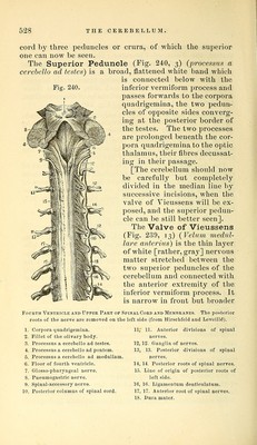
![THE CEREBELLUM. 52t still better seen], and between them is a thin layer of white matter, the Valve of Vieussens (Fig. 239, 13), to which the fourth pair of nerA^es may be traced round the superior peduncles of the cerebellum. The band of white matter passing transversely beneath the corpora quadrigemina on each side, and seen imme- diatelj^ in front of the superior peduncles of the cerebellum, is the FiUef of the Olimry body (Fig. 240, 2). Opportunity may now be taken to trace out the anterior commis- sure of the third ventricle and the anterior pillar of the fornix, by carefully scraping away the corpus striatum of one side. The Anterior Commissure is a cylindrical white band which may be traced through the corpus striatum to the roof of the descending cornu of the lateral ventricle. The Anterior pillar of the Fornix descends in front of the third ventricle and reaches the base of the brain, where it makes a twist to form the superficial white substance of the corpus albicans of one side, and then ascends to be lost in the gray matter of the optic thalamus (Figs. 237, p. 521, and 235). The Cerebellum. The Ceretellum (Fig. 231, 28, p. 501), or small brain, lies beneath the posterior lobes of the cerebrum, and in the skull is separated from them by the tentorium cere- belli. It is of a darker color than the cerebrum, and its surface is divided into laminae instead of convolutions, and these are separated by shallow sulci. The cerebellum is divisible into two lateral halves united by a commissure, and the horizontal fissure divides the organ into an upper and lower part. The upper surface is flat except in the median line, where there is a slight ridge forming the commissure, and called the superior verr^iiform process. The upper part of each hemisphere is divided into an anterior and a posterior lobe by an indistinct fissure. The anterior lohe is the larger and of a square shape, reaching as far back as the posterior extremity of the ver- miform process. The posterior lohe is the small portion behind the level of the vermiform process, and reaches to the horizontal fissure. The cerebellum is connected to the cerebrum and spinal](https://iiif.wellcomecollection.org/image/b21057679_0537.jp2/full/800%2C/0/default.jpg)