Practical anatomy: a manual of dissections / by Christopher Heath.
- Christopher Heath
- Date:
- 1870
Licence: Public Domain Mark
Credit: Practical anatomy: a manual of dissections / by Christopher Heath. Source: Wellcome Collection.
Provider: This material has been provided by the Francis A. Countway Library of Medicine, through the Medical Heritage Library. The original may be consulted at the Francis A. Countway Library of Medicine, Harvard Medical School.
539/600 (page 529)
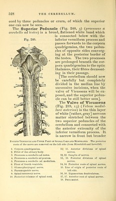
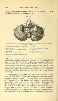
![behind, where it has a little gray matter connected with it, it is often torn through, in which case the cavity of the fourth ventricle is exposed. The fourth pair of nerves arises from the middle line of the valve close behind the corpora quadrigemina. The preparation is to be turned over for the examination of the remaining peduncles aud of the under surface of the cerebellum. The Middle Peduncle (Fig. 240, 4) processus a cere- hello ad pontem) has been already seen at the base of the brain. It is the largest of the three peduncles, and the fibres pass across the pons from one hemisphere of the cere- bellum to the other, constituting the [superficial] transverse fibres of the pons, which have been already dissected. The Inferior Peduncle (Fig. 240, 5) x>rocessus a cere- hello ad medullam) forms part of the restiform body of the medulla, and will be better seen when the fourth ventricle is opened. The Inferior Surface (Fig. 241) of the cerebellum is divided into two hemispheres by a deep fissure, the valle- cula, at the bottom of which is the inferior vermiform process, which is to be seen by drawing the medulla oblon- gata well forward. Each hemisphere is divided somewhat arbitrarily into lobes, since the divisions between them are very uncertain. Beginning behind is the PosteyHor Lohe (11); next the Slender Lohe (10) ; and in front of that the Biventral or Digastric Lohe (5). The Amygdala or Tonsil (Fig. 231, 29, p. 501) is a pro- minent lobe, close to the vallecula which it partially con- ceals. The Flocculus (Fig. 241, 4) is a small lobe immediately in front of the biventral lobe, which lies beneath the crus cerebelli, and is hence called the sub-peduncular lobe. The Inferior Vermiform Process (Fig. 241) is divided into the following portions. Most anteriorly is the Nodule (6), which projects into the fourth ventricle; posterior to this is the thin ridge called the Uvula (7) from the fact of its lying between the two tonsils ; behind this is the Pijramid (9), and posterior to this again are a few transverse commissural fibres. B}^ turning aside or cutting away the am3^gdalse, a laj'er of white matter will be brought into view, extending from 45](https://iiif.wellcomecollection.org/image/b21057679_0539.jp2/full/800%2C/0/default.jpg)