Practical anatomy: a manual of dissections / by Christopher Heath.
- Christopher Heath
- Date:
- 1870
Licence: Public Domain Mark
Credit: Practical anatomy: a manual of dissections / by Christopher Heath. Source: Wellcome Collection.
Provider: This material has been provided by the Francis A. Countway Library of Medicine, through the Medical Heritage Library. The original may be consulted at the Francis A. Countway Library of Medicine, Harvard Medical School.
543/600 (page 533)
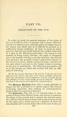
![PART YII. DISSECTION OF THE EYE. In order to study the general anatomy of the globe of the eye, it will be best to procure half a dozen bullocks' [sheep's or calves'] eyes, since the parts are larger than in the human eye, which also it is difficult to procure in a sufficiently recent condition. It must be borne in mind, however, that the eye of the bullock [&c.] differs from that of a man in the following particulars: the cornea is oval instead of being nearly circular; the pupil is elongated into a slit instead of being a circular aperture; the choroid coat presents the peculiar colored appearance known as the tapetum lucidum^ which is absent in man; and the yel- low spot which is present in the human retina is wanting in the eyes of quadrupeds. The following description will be of the human eye, which the student will find no diffi- culty in following. All the fat and the remnants of the muscles of the eye are to be removed with scissors, the optic nerve being carefully preserved. [The loose attachment of the conjunctiva over the sclerotic, and its firm adhesion over the corneal border, are to be observed. Also the insertion of the muscles just behind the corneal border.] The Human Eyeball (Fig. 243) is nearly globular in shape, but has a portion of a smaller sphere (the cornea) projecting anteriorly, thus making its antero-posterior greater than the transverse diameter. The Sclerotic (Fig. 243, ii) or external tunic is com- posed of dense white fibrous tissue, and serves to maintain the shape of the eyeball and to protect the internal parts. It is thicker behind than in front, and is pierced posteriorly by the optic nerve, which enters at a distance of about its own breadth [20°] to the inner side of the axis of the eye- 45*](https://iiif.wellcomecollection.org/image/b21057679_0543.jp2/full/800%2C/0/default.jpg)