Practical anatomy: a manual of dissections / by Christopher Heath.
- Christopher Heath
- Date:
- 1870
Licence: Public Domain Mark
Credit: Practical anatomy: a manual of dissections / by Christopher Heath. Source: Wellcome Collection.
Provider: This material has been provided by the Francis A. Countway Library of Medicine, through the Medical Heritage Library. The original may be consulted at the Francis A. Countway Library of Medicine, Harvard Medical School.
547/600 (page 537)
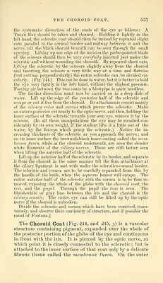
![tho systematic dissectiou of the coats of the eye as follows: A Third Eye should be taken and cleaned. Holding- it lightly in the left hand, the sclerotic coat should then be incised by repeated slight cuts parallel to the corneal border and midway between it and the nerve, till the black choroid beneath can be seen through the small opening. Lifting up one edge of the incision the probe-pointed blade of the scissors should then be very carefully inserted just under the sclerotic and without wounding the choroid. By repeated short cuts, lifting the sclerotic by the scissors slightly away from the choroid and inserting the scissors a very little way at a time and flatwise (but cutting perpendicularly) the entire sclerotic can be divided cir- cularly. (Fig. 244.) This can be done in water, but it is better to hold the eye very lightly in the left hand, without the slighest pressure. Forcing air between the two coats by a blowpipe is quite needless. The further dissection must now be carried on in a deep dish of ■water. Lift up the edge of the posterior half of the sclerotic and scrape or cut it free from the choroid. Its attachments consist mainly of the ciliary veins and nerves which pierce the sclerotic. Make an antero-posterior cut nearly to the optic nerve, and then, holding the inner surface of the sclerotic towards your own eye, remove it by the scissors. (In all these manipulations the eye may be steadied con- siderably by its own weight, if the student lifts it a little out of the water, by the forceps which grasp the sclerotic.) Notice the in- creasing thickness of the sclerotic as you approach the nerve ; and on its inner surface the brownish-black inner coating called the mem-. hrana fusca, while in the choroid underneath, are seen the slender white filaments of the ciliary nerves. These are still better seen when lifting the anterior half of the sclerotic. Lift up the anterior half of the sclerotic by its border, and separate it from the choroid in the same manner till the firm attachment at the ciliary ligament is met with under the sclerotico-corneal border. The sclerotic and cornea are to be carefully separated from this by the handle of the knife, when the aqueous humor will escape. The entire antei-ior half of the sclerotic with the cornea is to be thus re- moved, exposing the whole of the globe with the clioroid coat, the iris, and the pupil. Through the pupil the lens is seen. The bluish-white or gray line between the iris and the choroid is the ciliary muscle. The entire eye can still be lifted up by the optic nerve if the choroid is unbroken. Divide the sclerotic and cornea which have been removed, trans- versely, and observe their continuity of structure, and if possible the canal of Fontana.] The Choroid Coat (Fig. 244, and 245, 3) is a vascular structure containing pigment, expanded over the whole of the posterior portion of the globe of the eye and continuous in front with the iris. It is pierced by the optic nerve, at which point it is closely connected to the sclerotic; but is attached to the inner surface of that coat only by a delicate fibrous tissue called the membrana fusca. On the outer](https://iiif.wellcomecollection.org/image/b21057679_0547.jp2/full/800%2C/0/default.jpg)