The dissector's manual of practical and surgical anatomy / By Erasmus Wilson.
- Wilson, Erasmus, Sir, 1809-1884.
- Date:
- 1856
Licence: Public Domain Mark
Credit: The dissector's manual of practical and surgical anatomy / By Erasmus Wilson. Source: Wellcome Collection.
Provider: This material has been provided by the Harvey Cushing/John Hay Whitney Medical Library at Yale University, through the Medical Heritage Library. The original may be consulted at the Harvey Cushing/John Hay Whitney Medical Library at Yale University.
514/594 (page 520)
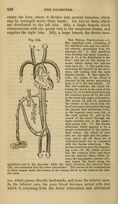
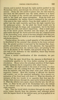
![enters the liver, where it divides into several branches, which may be arranged under three heads: 1st, two or three, which are distributed to the left lobe. 2dly, a single branch, which commnnicates with the portal vein in the transverse fissure, and supplies the right lobe. 3dly, a large branch, the ductus veno- ThE FfETAL CiRCtJLATIOy.—1. The umbilical cord, consisting of the umbilical vein and two umbili- cal arteries ; proceeding from the placenta (2). 3. The umbilical vein dividing into three branches ; two (4, 4), to be distributed to the liver ; and one (5), the ductus ve- nosus, which enters the inferior vena cava (6). 7, The portal vein, returning the blood from the intes- tines, and uniting with the right hepatic branch. 8. The right au- ricle ; the course of the blood is denoted by the arrow, proceeding from 8, to 9, the left auricle. 10. The left ventricle; the blood fol- lowing the arrow to the arch of the aorta (11), to be distributed through the branches given oflF by the arch to the head and upper extremities. The arrows 12 and 13, represent the return of the blood from the head and upper extremities through the jugular and subclavian veins, to the superior vena cava (14), to the right auricle (8), and in the course of the arrow through the right ventricle (15), to the pulmo- nar3'artery (16). 17. The ductus arteriosus, which appears to be a proper continuation of the pulmo- nary artery; the offsets at each side are the right and left pulmo- nary artery cut off; these are of extremely small size as compared with the ductus arteriosus. The ductus arteriosus joins the descend- ing aorta (IS, 18), which divides into the common iliacs, and these into the internal iliacs. which be- come the hypogastric arteries (19), and return the blood along the umbilical cord to the placenta; while the other divisions, the external iliacs (20), are continued into the lower extremities. The arrows at the terminations of these vessels mark the return of the venous blood by the veins to the infe- rior cava. \J I, siis, which passes directly backwards, and joins the inferior cava. In the inferior cava the ]iure blood becomes mixed with that which is returning from the lower extremities and abdominal](https://iiif.wellcomecollection.org/image/b20998831_0514.jp2/full/800%2C/0/default.jpg)