The dissector's manual of practical and surgical anatomy / By Erasmus Wilson.
- Wilson, Erasmus, Sir, 1809-1884.
- Date:
- 1856
Licence: Public Domain Mark
Credit: The dissector's manual of practical and surgical anatomy / By Erasmus Wilson. Source: Wellcome Collection.
Provider: This material has been provided by the Harvey Cushing/John Hay Whitney Medical Library at Yale University, through the Medical Heritage Library. The original may be consulted at the Harvey Cushing/John Hay Whitney Medical Library at Yale University.
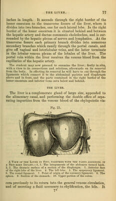
70/594 (page 76)
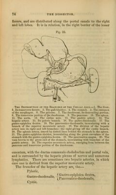
![the great curve of the stomach, l}Mri,2: between the layers of the p^reat omeiituin, and inosculates with the g'astro-epij>loica dextra. It is distributed to the greater curve of the stomach and to the great omentum. The ciASTRic VEINS, corresponding with the gastric, gastro- epiploic, and vasa brevia arteries, terminate in the splenic vein. The SPLENIC VEIN commences in the structure of the spleen, and quits that organ by several large veins; it is larger than the splenic artery, and perfectly straight in its course. It passes horizontally inwards behind the pancreas, and terminates near its greater end by uniting with the superior mesenteric and forming the portal vein. It receives in its course the gastric and pan- creatic veins, and near its termination the inferior mesenteric vein. The NERVES which accompany the branches of the coeliac axis are derived from the solar plexus, and constitute the gastric, hepatic, and splenic plexus. The relations of the vessels situated in the right border of the lesser omentum should now be examined more particularly. The hepatic artery will be found to the left, the ductus communis clioledoclius to the right, and the portal vein behind and between them. The student will also perceive how the lower boundary of the foramen of Wiuslow is formed by the hepatic artery. The ductus communis choledochns {xo'Kr-, bills, 6f;^o.uat, recipio) is the common excretory duct of the liver and gall-bladder. It is about three inches in length, and is formed by the junction of the hepatic with the cystic duct. It descends through the right border of the lesser omentum, and behind the descending por- tion of the duodenum to the inner side of that intestine, where it terminates by passing obliquely between the muscular and mu- cous coat, and opening on the summit of a papilla which is com- mon to it and the pancreatic duct. The ])a})illa is situated near the lower i)art of the descending j)ortion of the duodenum on its inner side ; and the duct is constricted in size during its passage between the coats of the intestine. The ductus communis choledochns, hepatic artery, and portal vein are surrounded and held to<,a»ther, while in the richt border of the k^ser omentum, by loose cellular tissue, which is continued with the vessels into the substance of the liver, and is termed (,'llssoirs capsule. In this Glisson's capsule are also contained a number of large lympliatic vessels which are taking tlieir course from the liver and gall-bladder to the lumbar glands. If the hepatic artery and ductus communis choledochns be drawn aside, and the connecting cellular tissue removed, the portal vein will be brought into view, lying between and behind the duct and artery. The VENA roinvE, formed by the union of the splenic and superior mesenteric vein behind the pancreas, is about three](https://iiif.wellcomecollection.org/image/b20998831_0070.jp2/full/800%2C/0/default.jpg)