The dissector's manual of practical and surgical anatomy / By Erasmus Wilson.
- Wilson, Erasmus, Sir, 1809-1884.
- Date:
- 1856
Licence: Public Domain Mark
Credit: The dissector's manual of practical and surgical anatomy / By Erasmus Wilson. Source: Wellcome Collection.
Provider: This material has been provided by the Harvey Cushing/John Hay Whitney Medical Library at Yale University, through the Medical Heritage Library. The original may be consulted at the Harvey Cushing/John Hay Whitney Medical Library at Yale University.
72/594 (page 78)
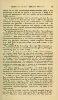
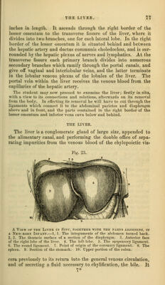
![is the largest organ in the body, weighing about four pounds, and measuring through its longest diameter about twelve inches. It is situated in the right hypochondriac region, and extends across the epigastrium into the left hypochondrium, frequently reaching, by its left extremity, the upper end of the spleen. It is placed obliquely in the abdomen ; its convex surface looking upwards and forwards, the concave downwards and backwards. The anterior border is sharp and free, and marked by a deep notch, the posterior rounded and broad. It is in relation, supe- riorly and posteriorly, with the diaphragm ; inferiorly, with the stomach, ascending portion of the duodenum, transverse colon, right supra-renal capsule, and right kidney; and corresponds, by its free border, with the lower margin of the ribs. Ligaments.—The liver is retained in its place by five liga- ments ; four of which are duplicatures of the peritoneum, situated on the convex surface of the organ ; the fifth is a fibrous cord which passes through a fissure in its under surface, from the um- bilicus to the inferior vena cava. They are, the— Longitudinal, Coronary, Two lateral, Round. The longitudinal ligament (broad, ligamentum suspensorium hepatis), is an antero-posterior fold of peritoneum, extending from the notch on the anterior margin of the liver to its poste- rior border. Between its two layers, in the anterior and free margin, is the round ligament. The lateral ligaments are formed by the two layers of perito- neum, which pass from the under surface of the diaphragm to the posterior border of the liver; they correspond with its lateral lobes. The coronary ligament \?, formed by the separation of the two layers forming the lateral ligaments near their point of con- vergence. The posterior layer is continued unbroken from one lateral ligament into the other ; but the anterior quits the pos- terior at each side, and is continuous with the corresponding layer of the longitudinal ligament. In this way a large oval surface on the posterior border of the liver is left uncovered by peritoneum, and is connected to the diaphragm by celhilar tissue. This space is formed principally by the right lateral ligament, and is pierced near its left extremity by the inferior vena cava, previously to the passage of that vessel through the tendinous oi)ening in the diaphragm. The round liganifnt is a fibrous cord resulting from the oblite- ration of the uinl)ilical vein, and situated between tlie two layers of })eritoneum in the anterior border of the h)ngitudina] ligament. It may be traced from the umbilicus, througli the longitudinal](https://iiif.wellcomecollection.org/image/b20998831_0072.jp2/full/800%2C/0/default.jpg)