The dissector's manual of practical and surgical anatomy / By Erasmus Wilson.
- Wilson, Erasmus, Sir, 1809-1884.
- Date:
- 1856
Licence: Public Domain Mark
Credit: The dissector's manual of practical and surgical anatomy / By Erasmus Wilson. Source: Wellcome Collection.
Provider: This material has been provided by the Harvey Cushing/John Hay Whitney Medical Library at Yale University, through the Medical Heritage Library. The original may be consulted at the Harvey Cushing/John Hay Whitney Medical Library at Yale University.
90/594 (page 96)
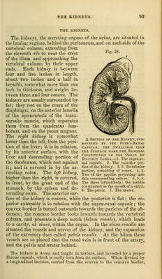
![that thej are the medium by which water, certain salts, and other sub- stances, pass out of the system; that they are, moreover, the means of escaj)e of certain morbid products, such as sugar, albumen, and the red particles of the blood. Respecting the cai)illary venous plexus, we have proof that the principal proximate constituents of urine, such as urea, lithic acid, &c., are, like the bile, derived from venous (portal) blood. The veins of the kidney commence at the surface by minute con- verging venules, the stelhited vessels, and proceed inwards, receiving in their course the veins of the cortical and tabular portions of the organ. On arriving at the pelvis, they unite to form the branches of the renal vein, which terminates in the vena cava by a single large trunk on each side; the left renal vein receiving the left spermatic vein. Injections thrown into the renal artery, and returning by the tubuli uriniferi, make their way into those tubes by rupture. The lymphatic vessels terminate in the lumbar glands. The nerves are derived from the renal plexus, which is formed partly by the solar plexus, and partly by the lesser splanchnic nerve. The renal plexus gives branches to the spermatic plexus, and branches which accompany the ureters: hence the morbid sympathies which exist be- tween the kidney, the iireter, and the testicle; and by the communica- tions with the solar plexus, with the stomach and diaphragm, and indeed witli the whole system. In the intimate structure of the kidney, the nerve-fibres are, according to Mr. Toynbee, continuous with the nucleated cells of the parenchyma of the organ. DEEP VESSELS AND NERVES OF THE ABDOMEN. The deep vessels and nerves of the abdomen are the abdominal aorta, inferior vena cava, thoracic duct, and sympathetic nerve. The duodenum and pancreas may now be removed, together with any cellular tissue, membrane, or organ which may imjx^de the view of the great vessels lying upon the vertebral column. In following the branches of the arteries and veins,*care should be taken to avoid destroying the nerves which lie upon the vessels and their numerous plexuses. The ABDOMiNx^L AORTA enters the abdomen through the aortic openinj^ of tlie diaphragm, and between the two ])illars of that muscle. In its course downwards, it lies on the left of the ver- tebral column, and terminates on the fourth luml)ar vertebra by dividing into the two common iliac arteries. It is crossed by the left renal vein, pancreas, transverse duodenum, and mesen- tery, and is in relation behind with the thoracic duct, recejita- culum chyli, and left lumbar veins. On its left side is situated the left semilunar ganglion and sympathetic nerve, and on its right, the inferior vena cava, right semilunar ganglion, and the commencement of the vena azygos. The branches of the abdomiiud aorta are, the— riirenic, Spermatic, f Gastric, Inferior mesenteric, Cieliac axis -f Hepatic, Siipra-rcnal, [Si)lenic, Kenal, Superior mesenteric, Lumbar, Sacra media.](https://iiif.wellcomecollection.org/image/b20998831_0090.jp2/full/800%2C/0/default.jpg)