Volume 1
Practical manual of diseases of women and uterine therapeutics : for students and practitioners / by H. Macnaughton-Jones.
- Macnaughton-Jones, H. (Henry), 1844-1918.
- Date:
- 1904
Licence: In copyright
Credit: Practical manual of diseases of women and uterine therapeutics : for students and practitioners / by H. Macnaughton-Jones. Source: Wellcome Collection.
97/712 (page 45)
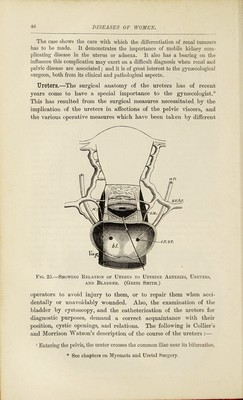
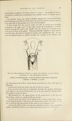
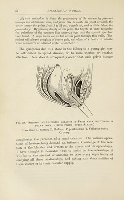
![diagnosing the disease, and advising an operation, or as surgeon in performing it. Only those who are frec^uently called upon to make a diagnosis can realize the difficulty there is in ariiving at an accurate conclusion in some obscure cases of renal disease if they be complicated with evidence of pelvic mischief, either remote or imme- diate. The vital imj)ortance of extreme care is obvious, as life may be sacrificed from the want of a simple exploratory incision, or the use of an aspirator. Mobile Kidney—Complicating Uterine Disease—Persistent High Temperature—Supervention of Carcinoma—Nephrectomy. The plates (II. and III.) show the right kidney removed by nephrectomy from a lady aged 40, one year after her first pregnancy. There had been a brown <lischarge from the uterus for a year, with cessation of the catamenia. During the entire year there was a constant nightly exacerbation of temperature, and uterine disease being suspected, slie was curetted. At the same time she l)ad an enlarged and movable kidney. Her symptoms not being relieved by curet- tage, exploration of the kidney was suggested. She had never had hsematuria, and there was nothing in the uterus indicative of malignant affection. The uterus and adnexa being healthy when I saw her, and the uterine tumour having increased in size, I suspected, from the emaciation and sickness, which were increasing, that the case was one of sarcoma. The kidiie}’- was removed by Langenbucli’s operation. She made an excellent recovery and put on flesh. Some eighteen months after the oi)eration disease recurred in the peri-renal tissue, and she died within two years from the primary operation.* Summary of the Pathological Report.—The specimen consists of an eidarged right kidney, weighing 2GJ ozs., and measuring 7 inches in length, and 11 inches in its greatest circumference. The enlargement is due to the presence of a new growth, which involves the lower two-thirds of the organ. This growth has a nodular surface, and is closely adherent to the fibrous capsule of the kidney, though it has not perforated the capsule. The hilum shows that the renal veins and pelvis are plugged 'with new growth. Tlie cut surface shows that the renal substance is entirely rejdaced by growdh at the lower end of the kidney. Microscopically, the growth is a very soft and degenerated carcinoma. Microscopical Report.—The growth itself is a carcinoma of the ‘ convoluted tube ’ type, that is to say, it reproduces the epithelium and general arrange- ment of the convoluted tubules more or less distinctly. Some of the alveoli have a lumen, and are even dilated into minute cysts, which present sim})le villous ingrowths or papillomata. The majority of the alveoli are, however, solid, and are separated by thin strands of fibrous tissue traversed by capillary vessels. A noteworthy feature of the growth is the marked fatty degenera- tion of the cells; this is shown by their empty, unstained condition, due to the I'emoval of the fat in the course of ])reparation of the specimen. * For particulars of case, see Brit. Gyn. Jour., Aug., 1897.](https://iiif.wellcomecollection.org/image/b28119605_0001_0097.jp2/full/800%2C/0/default.jpg)