Volume 1
On the anatomy, physiology, and pathology of the chimpanzee / by Charles F. Sonntag.
- Sonntag, Charles F. (Charles Frederick), -1925
- Date:
- 1923
Licence: In copyright
Credit: On the anatomy, physiology, and pathology of the chimpanzee / by Charles F. Sonntag. Source: Wellcome Collection.
34/118 page 354
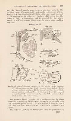
![processes. Its broad, flat tendon is attached to the ilio-pectineal line close to the emergence of the femoral vessels. The iliacus arises as in Man. It is quite continuous with the quadratus lumborum, and it soon fuses with the psoas magnus. its fibres envelop the psoas from each side. The psoas magnus blends more with the iliacus than in Man. It arises from the last dorsal vertebra, the inner inch of the last rib and the bodies and transverse processes of all the lumbar vertebrae. The com- bined muscle is inserted into the small trochanter and the femoral shaft a little below it. Half of the muscle (mesial part) passes over the. ilio-psoas tendon and is inserted into the bone posterior to the latter. The sartorius has a large fan-shaped aponeurotic origin from the anterior edge of the ilium up to a point a finger’s breadth below the anterior superior spine. Superficial to the aponeurosis, but connected to it by fascia, is a long, narrow fascial strip connecting the muscle to the anterior superior iliac spine. The strip is connected to the crural fascia and fascia over the abdominal muscles, thus forming a tunnel for the ilio-psoas. The comparatively slender muscle is inserted into the upper third of the anterior border of the tibia from the attachment of the ligamentum patellae downwards. Strong fascia unites it to the inner tuberosity of the tibia and the internal lateral ligament of the knee. Between it and the subjacent gracilis is the saphenous nerve, but no bursa. The gracilis and adductor longus (text-fig. 36, Gra. and Add. Long) arise by a common aponeurosis from the inner end of Poupart’s ligament, the entire length of the side of the symphysis, and upper third of the descending ramus of the pubis. No other author describes a precisely similar origin. And the origin conceals the adductor magnus. The adductor longus occupies the greater part of the aponeurosis, and its fibres approach closer to the bones ; it is inserted into the third quarter of the back of the shaft of the femur, and it is fused with the magnus. Hepburn (24) describes it as arising by a rounded tendon, and Humphry (26) gives its origin as the spine and inner half of the horizontal ramus of the pubis. The gracilis is inserted into the inner aspect of the tibia behind the internal lateral ligament. It is fused with the subjacent semitendinosus and the fascia over the inner head of the gastrocnemius. Gratiolet (22), Champneys (11), and Hepburn (24) give more extensive origins for a separate gracilis. The adductor magnus (text-fig. 36, Add.Mag) arises by three heads from the pubis and ischium, and it is inserted into the upper three-quarters of the back of the shaft of the femur. The upper head arises from the entire length of the body of the pubis : the middle head from the arch and ischial tuberosity: and the lower head from the tuberosity. The upper and mid heads unite to form a thick muscle inserted into the femoral shaft. The lower head runs separately to the adductor tubercle, [32]](https://iiif.wellcomecollection.org/image/b2982123x_0001_0034.jp2/full/800%2C/0/default.jpg)