Volume 1
On the anatomy, physiology, and pathology of the chimpanzee / by Charles F. Sonntag.
- Sonntag, Charles F. (Charles Frederick), -1925
- Date:
- 1923
Licence: In copyright
Credit: On the anatomy, physiology, and pathology of the chimpanzee / by Charles F. Sonntag. Source: Wellcome Collection.
35/118 page 355
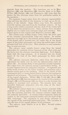
![and the femoral vessels pass between the two parts to the popliteal space. Ohampneys (11) reviews the various descriptions. Dwight (10) describes fusion of all the adductors except the part of the magnus to the tubercle. Hepburn (24) states that the latter is really a hamstring, and is supplied by the sciatic nerve. I did not observe fibres from the main mass reaching the knee. Text-figure 36. ADD. LONG. Muscles and joints of the pelvic extremity. A.C.L: anterior cruciate ligament; C.T : fibrous connecting slip; E.L.H : extensor lougus liallucis; E.S.C: external semilunar cartilage; I.C.B: internal cuneiform bone; I.S.C : internal semilunar cartilage; Met.I : first metatarsal bone; N.B : scaphoid or navicular; P.C.L : posterior cruciate ligament; PYR : pyriformis ; T. A : tibialis anticus. Other letters in text. The adductor brevis (text-fig. 36, Add.Brev) arises by three prismatic, interlocking bellies from the angle between the body and horizontal pubic ramus. Its flat tendon is inserted into a line from the small trochanter to the middle of the back of the femur. The pectineus (text-fig. 36, Pect) arises from the pubic crest on [33] ,](https://iiif.wellcomecollection.org/image/b2982123x_0001_0035.jp2/full/800%2C/0/default.jpg)