Volume 1
On the anatomy, physiology, and pathology of the chimpanzee / by Charles F. Sonntag.
- Sonntag, Charles F. (Charles Frederick), -1925
- Date:
- 1923
Licence: In copyright
Credit: On the anatomy, physiology, and pathology of the chimpanzee / by Charles F. Sonntag. Source: Wellcome Collection.
36/118 page 356
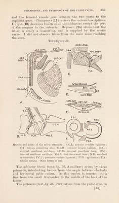
![the three-quarters of an inch internal to the longus. It has a curved insertion running from the lesser trochanter to the back of the shaft of the femur. Hamstring Muscles:—With the exception of the short head of the biceps, all the hamstrings arise together from the lower and back part of the tuber ischii, and all are fused. The semitendi- nosus is inserted into the anterior tubercle of the tibia, and by a large expansion to the fascia of the leg. The tendon is not as long as in Man (Vrolik and Hepburn), and the insertion is lower. Cuvier showed that this low insertion is incompatible with an erect attitude, and Rolleston pointed out that it occurs in children. The insertion of this muscle, and that of the semimembranosus move upwards as the body becomes erect. The semimembranosus is smaller than the last muscle, and its long, flat tendon sends no fibres to the fascia over the popliteus, nor to the internal lateral ligament. It is inserted into the tibia over a small area proximal to the other hamstrings. The biceps differs somewhat in my specimen from the accounts of Hepburn (24) and Champneys (11). Both heads remain separate. The ischial head has a strong insertion into the outer side of the head of the tibia, the head of the fibula, and the fascia over the outer head of the gastrocnemius. The femoral head is inserted into the head and proximal inch of the shaft of the fibula, and the fascia over the gastrocnemius. It is, therefore, evident that the hamstrings and some of the adductor group are connected to the fascia over the gastrocnemius. The gluteus maximus is smaller than in Man. It arises from the side of the sacrum and coccyx, the great sacro-sciatic ligament and the ischial tuberosity along with the long head of the biceps. No muscle fibres arise from the iliac crest, as in Phascolarctos, but it arises from the strong fascia which covers the gluteus medius* and is attached to the iliac crest. Hepburn (24) saw it arising from the crest, Humphry (26) observed no fascial origin, and Champneys (11) described conditions similar to mine. The insertion is longer than in Man, for it is fixed to the back of the femur as low down as the external condyle, and to the shaft below the great trochanter. Its fibres mingle with the vastus externus. It is fused with the outer head of the gastro- cnemius (Humphry, 26), and with the tensor fasciae femoris (Wilder, 53). The gluteus medius has a fleshy origin from the whole of the dorsum ilii down to the line from the great sciatic notch to the anterior inferior spine, and by a dense aponeurosis from the anterior border between the superior and inferior spines. The aponeurosis gives way to muscle after an inch. Fibres also arise from the posterior aspect of the aponeurosis. It is inserted into the top of the outer aspect of the great trochanter. A small slip runs from the mesial aspect of the tendon to join the tendon of the pyriformis, thus bringing the two tendons into connection. Two communicating bursae (text-fig. 36, Bur) separate the [34]](https://iiif.wellcomecollection.org/image/b2982123x_0001_0036.jp2/full/800%2C/0/default.jpg)