Volume 1
On the anatomy, physiology, and pathology of the chimpanzee / by Charles F. Sonntag.
- Sonntag, Charles F. (Charles Frederick), -1925
- Date:
- 1923
Licence: In copyright
Credit: On the anatomy, physiology, and pathology of the chimpanzee / by Charles F. Sonntag. Source: Wellcome Collection.
44/118 page 364
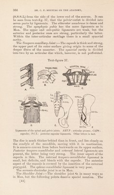
![The Joints. Spinal Ligaments :—The anterior common ligament extends from the axis to the upper segment of the sacrum, and is smaller above than below. It is attached to the front of the centra, but not to the depressions between the vertebrae. The supraspinous ligaments run as in Man, and are connected to interspinous ligaments. There is no ligamentum nuchae. The interspinous ligaments are as in Man. The ligamenta subjlava run from the anterior aspect of the lamina above to the posterior aspect of the lamina below. They and the interspinous ligaments are very elastic. The posterior common ligament runs from the axis to the sacrum, but is not so dentate in appearance as in Man. It is narrow on the centra, and expanded over the intervertebral discs. Costal Articulations (text-fig. 37 A & B):—Anteriorly the head of the rib is connected by a fan-shaped ligament, not divisible into three parts as it is in Man, and the fibres all gain attachments to the anterior common ligament (A.C.L). The upper fibres run to the vertebra above, and the lower ones to the vertebra below, where they are overlapped by the upper fibres of the next joint. Posteriorly the tubercle of the rib is attached to the transverse process of the vertebra by a capsule. The upper border of the neck of the rib is connected to the transverse process by a fan-shaped ligament (C-T.L.). The intertransverse ligaments (I-T.L) are fan-shaped. They connect the tip of the transverse process above to the upper border of the transverse process below. The lengths of the spinous processes of the cervical vertebrae are:—axial | inch ; 4th vertebra | inch; 6th and 7th vertebrae g inch. Atlanto-axoid Joints :—The posterior a/tlanto-axoid ligament, from the posterior arch of the atlas to the upper border of the axis, corresponds to the ligamenta subflava elsewhere. It is strengthened by fibrous bands (text-fig. 37, F.B) : a. From the transverse process of the axis to an elevation on the posterior arch of the atlas; b. A central atlanto-axoid band (C.A.B); c. Several small bands between the others. The anterior atlanto- axoicl ligament is a very strong band continuing the anterior common ligament from the axis to the atlas. Joints of the Occipital, Atlas, and Axis (text-fig. 37 I)):—The posterior occipito-atlantoid ligament is a thin membrane running between the posterior arch of the atlas and the edge of the foramen magnum. It is strengthened by lateral bands, attached close to the cond}des. It functions as a posterior capsular liga- ment. An internal capsular ligament runs from the inner border of the condyle to the superior articular process of the atlas. And a strengthening band runs from the posterior arch of the atlas to the occipital bone close to the attachment of the internal capsular ligament. The anterior occipito-atlantoid [42]](https://iiif.wellcomecollection.org/image/b2982123x_0001_0044.jp2/full/800%2C/0/default.jpg)