Volume 1
On the anatomy, physiology, and pathology of the chimpanzee / by Charles F. Sonntag.
- Sonntag, Charles F. (Charles Frederick), -1925
- Date:
- 1923
Licence: In copyright
Credit: On the anatomy, physiology, and pathology of the chimpanzee / by Charles F. Sonntag. Source: Wellcome Collection.
5/118 page 325
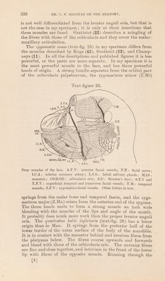
![A fan-slm.ped muscle separates from the platysma in the neck, runs upwards behind the auricle and spreads out into bundles which are attached to the back of the auricle, the occipital crest, occipitalis muscle, and the deep fascia over the back of the neck. Huge (42) has given a very elaborate account of the manner in which the platysma enters into the other facial muscles. Occipito-frontalis (text-fig. 25):—There are many differences of opinion about this muscle. Tyson (50) and Traill (49) could not detect it, Owen (39) found a trace of it, and Wilder (53) found the muscle bellies small, but the aponeurosis was large. Huge (42) figured a very extensive muscle and a small aponeurosis. In my specimen the occipitalis arises from the middle two- fourths of the occipital crest, but it is not divisible into two bellies as in Man. The fibres pass forwards for nearly two inches and end in a well-marked aponeurosis. The frontalis arises from the supra-orbital ridges and space between, but is not so well- marked as the occipitalis. It is very easily removed with the skin. It blends with the orbicularis oculi. The Orbicularis oculi (text-fig. 27 A) is divisible into orbital and palpebral parts as in Man. The former arises from the inner end of the frontal bone and the nasal process of the maxilla; and both muscles are united across the mid line. As it lies on the bones bounding the orbit its upper part is strong and compact and gives offa strong bundle of fibres from its lateral part to enter the zygomatic mass (Z.M). The fibres on the lower boundary of the orbit are arranged in loose bundles. The palpebral fibres run from the internal tarsal ligament to the lateral tarsal raphe, and are thickened close to the roots of the eyelashes, the thickened parts being of greater dimensions than the ciliary bundles (O.B) in Man. At the lateral tarsal raphe the orbital and palpebral parts are continuous. The nerve-supply from the facial nerve is shown in text-fig. 26. The lips and cheeks' receive many muscles (text-fig. 25), most of which, though thin, are of considerable superficial extent. They are disposed in two layers as in Man, but the characters are very different in a number of points. The superficial layer, is composed of the risorius, levator labii superioris, zygomatic mass, orbicularis oris, triangularis and quadratus labii inferioris. The deep layer consists of buccinator, depressor anguli oris, incisivi, canini, mentales, and premolares. The risorius is com- posed entirely of the upper part of the platysma, for no fibres are derived from the fascia over the masseter muscle. It blends with other muscles at the angle of the mouth. The levator labii superioris (Lev. Lab. Sup) arises, under cover of the orbital part of the orbicularis oculi, from the entire infra-orbital border of the maxilla. It radiates in a fan-like manner and is inserted into the entire length of the upper lip and upper border of the alge nasi. The fibres forming the latter insertion correspond to the levator labii superioris alseque nasi of Man. Many of the fibres of the muscle are very thin. Champneys (11) states that it Proc. Zool. Soc.—1923, No. XXII. 22 [3]](https://iiif.wellcomecollection.org/image/b2982123x_0001_0005.jp2/full/800%2C/0/default.jpg)