Volume 1
On the anatomy, physiology, and pathology of the chimpanzee / by Charles F. Sonntag.
- Sonntag, Charles F. (Charles Frederick), -1925
- Date:
- 1923
Licence: In copyright
Credit: On the anatomy, physiology, and pathology of the chimpanzee / by Charles F. Sonntag. Source: Wellcome Collection.
50/118 page 370
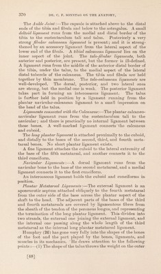
![The Ankle Joint:—The capsnle is attached above to the distal ends of the tibia and fibula and below to the astragalus. A small deltoid ligament runs from the medial and distal border of the tibia to the sustentaculum tali and talus. Posteriorly a very strong Jlbular calcanean ligament is present; and it is streng- thened by an accessory ligament from the lateral aspect of the lower end of the fibula. A tibial calcanean ligament lies on the inner aspect of the joint. The talo-fibular ligaments, both anterior and posterior, are present, but the former is ill-defined. A ligament runs from the middle of the anterior distal border of the tibia, under the talus, to the medial aspect of the lateral distal tubercle of the calcaneus. The tibia and fibula are held together by thin membrane. The talo-calcanean ligaments are well-developed. The dorsal, posterior, and two lateral ones are strong, but the medial one is weak. The posterior ligament takes part in forming an interosseous ligament. The talus is farther held in position by a ligament running from the plantar navicular-calcanean ligament to a small impression on the head of the talus. Ligaments connected with the Calcaneus'.—The plantar calcaneo- navicular ligament runs from the sustentaculum tali to the navicular; and there is practically no internal ligament between these bones. A well-marked ligament connects the calcaneus and cuboid. The long plantar ligament is attached proximally to the cuboid, and distally to the bases of the second, third, and fourth meta- tarsal bones. No short plantar ligament exists. A fine ligament attaches the cuboid to the lateral extremity of the base of the fifth metatarsal, and another connects it to the third cuneiform. Navicular Ligaments:—A dorsal ligament runs from the navicular bone to the base of the second metatarsal, and a medial ligament connects it to the first cuneiform. An interosseous ligament holds the cuboid and cuneiforms in position. Plantar Metatarsal Ligaments:—The external ligament is an aponeurotic septum attached obliquely to the fourth metatarsal from the outer side of the base across the plantar aspect of the shaft to the head. The adjacent parts of the bases of the third and fourth metatarsals are covered by ligamentous fibres from the sheath of the tendon of the peroneus longus, and representing the termination of the long plantar ligament. This divides into two strands, the external one joining the external ligament, and the internal one passing along the whole length of the third metatarsal as the internal long plantar metatarsal ligament. Humphry (26) has gone very fully into the shapes of the bones of the foot and the part played by the bones, ligaments, and muscles in its mechanics. He draws attention to the following points :—(1) The shape of the talus throws the weight on the outer [48]](https://iiif.wellcomecollection.org/image/b2982123x_0001_0050.jp2/full/800%2C/0/default.jpg)


 No text description is available for this image
 No text description is available for this image
 No text description is available for this image