Volume 1
On the anatomy, physiology, and pathology of the chimpanzee / by Charles F. Sonntag.
- Sonntag, Charles F. (Charles Frederick), -1925
- Date:
- 1923
Licence: In copyright
Credit: On the anatomy, physiology, and pathology of the chimpanzee / by Charles F. Sonntag. Source: Wellcome Collection.
62/118 page 382
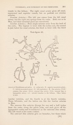
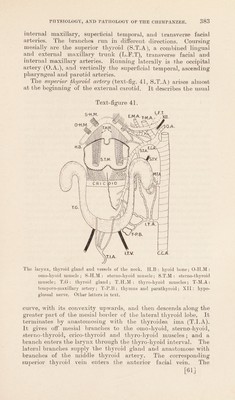
![arise from them in Man are here replaced by branches of the last lumbar and femoral arteries. The relations are as in Man. The hypogastric artery (text-fig. 40 E) on each side divides as in Man into anterior and posterior divisions. The anterior division gives off:— 1. A trunk which divides into superior vesical (S.V.A) and uteri-vaginal (U-V.A) arteries. The former is a small vessel, which reaches the side of the bladder up which it runs to the fundus. The latter is larger and breaks up into vessels supplying the uterus and vagina ; details are given on p. 401. 2. The Pudendal Artery (P.A) gives off the inferior vesical artery (I.V.A), passes through the great sciatic notch at the lower border of the pyriformis and, after giving off the inferior gluteal artery (I.G.A), it breaks up into branches which pass through the ischio-rectal fossa to the rectum and anus, the vagina, the levator ani and sphincter vaginse. The posterior division gives off:— 1. A bundle of arteries which enter the anterior sacral foramina (S.A.). 2. Lateral sacral artery (L.S.A). 3. Superior gluteal artery (S.G.A). The superior gluteal artery emerges at the upper border of the pyriformis and supplies it. It divides into two main branches. The upper one supplies the gluteus medius and gives the nutrient artery to the ilium. The lower one descends to supply the gluteus medius, gluteus minimus, and scansorius. The inferior gluteal artery emerges with the pudendal artery at the lower border of the pyriformis. It gives branches to the gluteus maximus, gemellus superior, obturator interims, scan- sorius, and acetabular part of the ilium. It anastomoses with the lateral circumflex artery. Arteries of the Head and Neck. The common carotid arteries {text-fig. 41,C.C.A) extend from the sterno-clavicular articulations to the upper border of the lateral aspects of the thyroid cartilage, where they divide into external and internal carotids. They are concealed by the large external jugular veins, and they line in front of the vagus and sympathetic nerves. But no internal jugular veins are present to form lateral relations. No carotid sheath exists. The other relations are as in Man. It gives off tortuous inferior and middle thyroid arteries (text-fig. 41, I.T.A. and M.T.A), which replace the inferior thyroid branch of the subclavian arteries. This may be an individual peculiarity. The external carotid artery (E.C.A) first ascends almost verti- cally till it reaches the level of the hyoid bone, where it inclines posteriorly and upwards, being continued as the temporo- maxillary artery. Within the parotid gland it divides into [60]](https://iiif.wellcomecollection.org/image/b2982123x_0001_0062.jp2/full/800%2C/0/default.jpg)
 No text description is available for this image
 No text description is available for this image
 No text description is available for this image