Volume 1
On the anatomy, physiology, and pathology of the chimpanzee / by Charles F. Sonntag.
- Sonntag, Charles F. (Charles Frederick), -1925
- Date:
- 1923
Licence: In copyright
Credit: On the anatomy, physiology, and pathology of the chimpanzee / by Charles F. Sonntag. Source: Wellcome Collection.
8/118 page 328
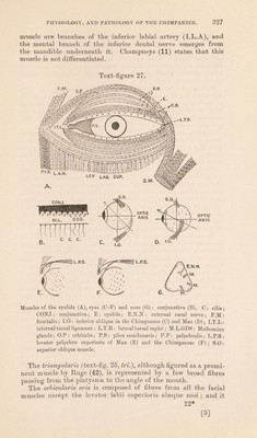
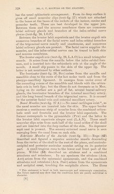
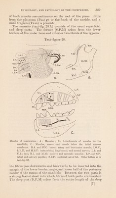
![has the usual sphincteric arrangement. From its deep surface it gives off small muscular slips (text-fig. 27) which are attached to the bones at the bases of the sockets of the incisor, canine and premolar teeth. These are best developed in the upper jaw. Between them and the mucous membrane there are numerous labial salivary glands and branches of the infra-orbital nerve plexus (text-fig. 26, I.O.P). Between the levator labii superioris and the levator anguli oris numerous branches of the facial nerve and infra-orbital branches of the trigeminal nerve ramify and anastomose, and numerous labial salivary glands are present. The facial nerve supplies the muscles, and the infra-orbital nerves can be traced to both skin and mucous membrane. The levator anguli oris (text-fig. 26, L.A.O) is a small triangular muscle. It arises from the maxilla below the infra-orbital fora- men, and is inserted into the orbicularis oris at the angle of the mouth. A small slip passes to the skin of the upper lip. The latter is not mentioned by other authors. The buccinator (text-fig. 26, Buc) arises from the maxilla and mandible close to the roots of the last molar teeth and from the pterygo-maxillary ligament. It emerges from under cover of the ascending ramus of the mandible and blends with the orbicu- laris oris in both lips; but the fibres do not decussate as in Man. Lying on its surface are a pad of fat, several buccal salivary glands, the buccinator branches of the internal maxillary artery, and the long buccal branch of the trigeminal nerve. It is crossed by the anterior facial vein and external maxillary artery. Nasal Muscles (text-fig. 27 A) :—No nasal cartilages exist*, so the nasal muscles are inserted into the skin. The upper border receives a continuous strip of muscles from the combined orbicu- laris oculi and frontalis and the levator labii superioris. The former corresponds to the pyramidalis (Pyr) and the latter to the levator labii superioris alseque nasi (L.A.N). Three small muscular slips arise from each half of the nasal orifice of the skull and are inserted into the deep surface of the skin. No depressor septi nasi is present. The sensory external nasal nerve is seen emerging from the nasal fossa on each side. Extrinsic Muscles of the Auricle (text-fig. 25):—Huge (42) 'described and figured a superior auricular muscle descending from the vertex of the skull to the root of the pinna., and auriculo- occipital and posterior auricular muscles acting on its posterior j^art. A small tragicus runs to the lowTer and front part of the pinna. Wilder (53) described an attolens and a combined attrahens and retraliens. In my specimen the attolens (Aur. Ant) arises from the epicranial aponeurosis, and the combined attrahens and retrahens (Aur. Post) arises from the aponeurosis and occipital crest, touching the occipitalis above. The fibres * This statement is based on both macroscopic and microscopic examination. But future material may show that the conditions here are purely individual in character. [6]](https://iiif.wellcomecollection.org/image/b2982123x_0001_0008.jp2/full/800%2C/0/default.jpg)
 No text description is available for this image
 No text description is available for this image
 No text description is available for this image