Volume 1
On the anatomy, physiology, and pathology of the chimpanzee / by Charles F. Sonntag.
- Sonntag, Charles F. (Charles Frederick), -1925
- Date:
- 1923
Licence: In copyright
Credit: On the anatomy, physiology, and pathology of the chimpanzee / by Charles F. Sonntag. Source: Wellcome Collection.
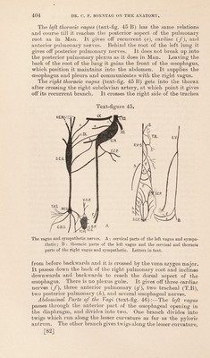
81/118 page 401
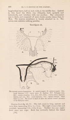
![ligament at a higher level than the uterus. The histology has been described by Giacomini (66), Duval (67), and Sperino (47). Ligaments connect it to the utero-tubal junction, and to the tube below and behind the fimbriae. The ovarian fimbria is well marked. Sperino describes triangular ovaria. The primordial ova are innumerable, and are similar to those in the human species. And Graafian follicles can be seen in various stages of development according to Sperino. Fallopian Tubes:—Both are 6‘5cm. long when drawn straight. They hardly increase in calibre from their uterine to their ovarian ends. The fimbriae form a dense cluster, the ovarian fimbria is well marked, and uterine and abdominal orifices are plain; but one cannot easily pass a bristle through the tube. Each tube curves over the anterior border and upper pole of the corresponding ovary. The hydatid (text-fig. 44, H.M) is well marked on the right side. The epoophoron and paroophoron are present. Uterus (text-fig. 44):—The uterus is isolated from the bladder and rectum by peritoneal fossae, and its summit lies L5 cm. above the floor of each. There is no marked fundus, the body is triangular and the cervix is fusiform. The body is P5 cm. long, and its base is L5 cm. across. The cervix is P2 cm. long, and 1*1 cm. across at its widest part. It has very infantile propor- tions. The round ligaments are large and run directly upwards and forwards from the utero-tubal junction. The interior of the body of the uterus is smooth between the tubes, but lower down it has an upward continuation of the median dorsal crest and transverse ridges which occupy the cervix. The musculature in the upper part of the uterus is thinner than in the lower part of the body and the cervix. The external os uteri is oval, with nodulated continuous lips. Both lips are of equal length. This account differs in several respects from the accounts of Sperino (47), and others. Gratiolet (22) described a bicornuate uterus. The vagina is 5 cm. long, and expands from above downwards. Anterior and posterior fornices are both present, but the latter is much the larger. In its upper part there is a median dorsal cushion, and the mucosa has transverse folds. Below that it has longitudinal folds. In its lower part it has fine longitudinal striae and several pockets (text-fig. 44). The urethra opens on its anterior wall about the middle. The uterine artery (U.A) supplies the vagina, uterus, tubes, ovaries, epoophoron, etc. It anastomoses with the very small ovarian artery. Its complexity is shown in text-fig. 44. The external generative organs (Plate I. B) are built on the same plan as, but differ from those of the human female. The mons veneris (M.Y) is slight, and has a few hairs. The labia majora (L.M) are represented by slight elevations of skin over thick- enings of the subcutaneous fat. The labia minora (L.Mi) are large and folded, and divide to surround the large clitoris (CL) the latter having two crura covered by well-developed ischio- cavernosi muscles. A small fourchette exists, but there is no [79]](https://iiif.wellcomecollection.org/image/b2982123x_0001_0081.jp2/full/800%2C/0/default.jpg)