Volume 1
On the anatomy, physiology, and pathology of the chimpanzee / by Charles F. Sonntag.
- Sonntag, Charles F. (Charles Frederick), -1925
- Date:
- 1923
Licence: In copyright
Credit: On the anatomy, physiology, and pathology of the chimpanzee / by Charles F. Sonntag. Source: Wellcome Collection.
82/118 page 402
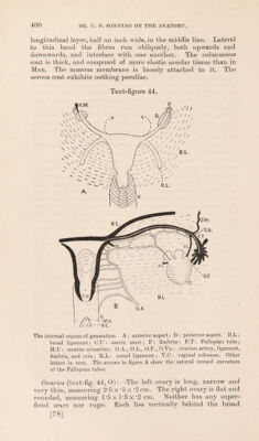
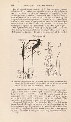
![hymen. The meatus urinarius is within the vagina, so no prominent vestibule is seen as in the human condition. The glands of Bartholin lie between the vagina and rectum. Sperino (47), Bischoff (60), Chapman (12), Gratiolet (22), Hartmann (65), Barkow (2), Hoffmann (68), Symington (48), and Traill (49) have described the external genitalia; and many of these ob- servers have described the internal organs. Winwoode Beade(57), Garner (21), and Mohrike (35) describe a sexual season, and Bolau (5), Elders (59), Hermes (69), and Keith (30) describe either the periodicity or characters of men- struation. Pocock (80) contrasts menstruation in the Chim- panzee and Hainan Gibbon. The Nervous System #. The olfactory nerve terminates by marked branches on the upper thirds of the turbinate regions and nasal septum. The optic nerve is large and surrounded by a sheath of dura mater. No arteria retinae centralis was detected in it, but the injection material may not have traversed it. The oculo-motor nerve has superior and inferior divisions. The superior division does not pierce, but runs to the inner side of, the superior rectus. It supplies the superior and internal rectus muscles and ends in the levator palpebrae superioris. The inferior division runs downwards and outwards on the outer side of the rectus inferior, gives a motor branch to the ciliary ganglion, supplies the inferior rectus and ends in the inferior oblique. The branch of the superior division to the internal rectus is very large. The trochlear nerve ends by three divisions to the superior oblique muscle. The trigeminal nerve has three divisions as in Man, radiating from the Gasserian ganglion. The ophthalmic division courses as in Man, and breaks up into:—1. Lachrymal nerve, lying between the orbital wall and upper border of the external rectus. It supplies the lachrymal gland, conjunctiva and skin of the eyelids. 2. Frontal nerve resembles that in Man. It breaks up into supra-orbital and supra-trochlear branches. 3. Nasal nerve. This is distributed as in Man, but the lateral terminal branch, which is very large, comes out of the nasal cavity direct, and not between bone and cartilage, as in Man. The ciliary ganglion is larger than in Man. It lies on the lateral side of the oph- thalmic artery and receives filaments from both divisions of the third nerve, a twig from the naso-ciliary nerve, and sympathetic filaments from the carotid plexus. It gives off short ciliary nerves: one large one, lying on the outer side of the optic nerve, divides into upper and lower divisions on reaching the eyeball. The superior and inferior maxillary divisions of the trigeminal are similar to those in Man, but I was unable to detect as many branches radiating from Meckel's ganglion. The chorda tympani * The brain will be described in a separate paper by Professor G. Elliot Smith, F.R.S. [80]](https://iiif.wellcomecollection.org/image/b2982123x_0001_0082.jp2/full/800%2C/0/default.jpg)