Volume 1
On the anatomy, physiology, and pathology of the chimpanzee / by Charles F. Sonntag.
- Sonntag, Charles F. (Charles Frederick), -1925
- Date:
- 1923
Licence: In copyright
Credit: On the anatomy, physiology, and pathology of the chimpanzee / by Charles F. Sonntag. Source: Wellcome Collection.
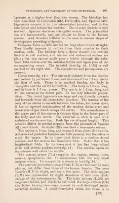
83/118 page 403
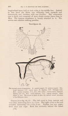
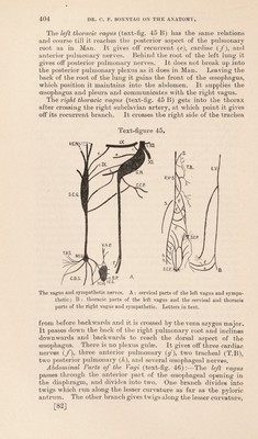
![joins the inferior maxillary division before the latter separates into its lingual and inferior dental nerves. The submaxillary ganglion is not separate as in Man, but is fused with the hypoglossal nerve. The otic ganglion was not recognised with certainty. The abducens emerges between the two heads of the external rectus and sinks into the ocular surface of the muscle. The facial nerve emerges from the stylo-mastoid foramen. Its intra-petrous course was not traced. It divides in the parotid gland into temporal, zygomatic, maxillary, buccal, mandibular, and cervical divisions. The temporal branches run upwards and are distributed as in Man. The zygomatic and maxillary divisions eventually unite and give off from their combined trunk a number of branches to the muscles of the face. The mandibular and cervical divisions are as in Man. The union of the chorda tympani and trigeminal nerves has already been described. The auditory nerve was not traced. The glosso-pharyngeal nerve emerges from the inner part of the jugular foramen and communicates with the other nerves at the upper part of the neck. It passes between the external and internal carotid arteries, curves round the stylo-pharyngeus muscle and disappears under the free outer edge of the hyoglossus. Finally it breaks up into branches to the tongue, pharynx, and tonsil. It supplies the stylo-pharyngeus. The tympanic and petrosal branches were not traced. The Vagus Nerve (text-figs, 45 h 46) emerges from the jugular foramen wherein it is lateral to the glosso-pharyngeal nerve, posterior to the internal jugular vein and mesial to the accessory nerve, to which it is closely adherent. Immediately below the base of the skull it develops the ganglion nodosum (G.N.) on its lateral aspect. The nerve separates from the ganglion again at the level of the posterior border of the hard palate. At the root of the neck it runs on to the posterior aspect of the common carotid artery and then it enters the thorax on the left side. The right one disappears under cover of the innominate artery where the latter bifurcates into right common carotid and sub- clavian arteries. The left vagus (text-fig. 45 A) only communicates with the sympathetic, but the right one (text-fig. 45 B) is ex- tensively used with the sympathetic. Branches in the Neck :— 1. Communicating nerves to the glosso-pharyngeal (c. ix), hypoglossal (c. xii), superior cervical ganglion of the sympathetic (S.C.G) and cervical plexus (c.C.P). 2. Pharyngeal nerve (a). 3. Superior laryngeal nerve (b). 4. Bight recurrent laryngeal nerve (d). 5. Cardiac branch of the left vagus (f). 6. Plexus of carotid, tracheal and cardiac branches of the right vagus. [81]](https://iiif.wellcomecollection.org/image/b2982123x_0001_0083.jp2/full/800%2C/0/default.jpg)