Volume 1
On the anatomy, physiology, and pathology of the chimpanzee / by Charles F. Sonntag.
- Sonntag, Charles F. (Charles Frederick), -1925
- Date:
- 1923
Licence: In copyright
Credit: On the anatomy, physiology, and pathology of the chimpanzee / by Charles F. Sonntag. Source: Wellcome Collection.
93/118 page 413
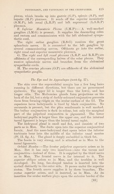
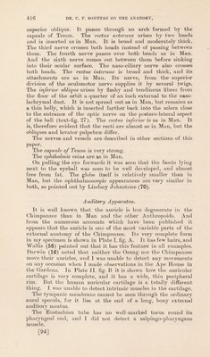
![The Sympathetic Nervous System (text-figs. 45 & 46). The long, oval superior cervical ganglion (S.C.G) extends from the level of the hard palate to the hyoid bone. It is connected by communicating branches to the ninth (IX) and twelfth cranial nerves, and to the ganglion nodosum (G.N) and its superior laryngeal (b) branch. On the left side it sends no twigs direct to the cervical plexus, but it is connected to the first and second cervical nerves on the right (text-fig. 45 A.) It gives off pharyngeal nerves and the external carotid plexus, but no cardiac nerve arises from it. The internal carotid branch (I.G.N) breaks up into a plexus before it enters the skull. The left sympathetic runs separate from the vagus and ends in the middle cervical ganglion (M.C.G) whence the following branches radiate:—(1) A stout cord which divides into branches accompanying the thyroidea ima artery to the thyroid gland (T.B.S), tracheal nerves and cardiac nerves (C.B.S) to the deep part of the cardiac plexus and plexus round branches of the aortic arch. (2) Nerves to the cardiac and aortic plexus (C.B.S) (3) Continuation of the cord to the inferior cervical ganglion (I.C.G). This also communicates with the vertebral plexus (V.A.P), brachial plexus (c.B.P), and cardiac plexus. The right sympathetic fuses with the right vagus, but separates from it lower down again, and a rich plexus of nerves comes from it, both above and below, and accompanies the common carotid artery to the plexus on the branches of the aortic arch. The middle ganglion does not send off* many radiations as on the left side. The inferior cervical ganglion (I.C.G) and first thoracic ganglia are fused. It gives off' rami communicantes to the brachial plexus (c.B.P), a thick plexus which accompanies the vertebral artery (V.A.P), a nerve to the cardiac plexus, and the thoracic sympathetic cord (T.C.S). The Thoracic Cords have fewer ganglia than the number of intercostal nerves. The left one gives off the great splanchnic nerve (G.S.N) at the level of the fifth and sixth thoracic nerves. At the level of the diaphragm it divides into the small splanchnic nerve (S.S.N) and abdominal sympathetic cord (S.C). In addition to these it gives off rami communicantes to the intercostal nerves and some of these are long. Aortic nerves accompany the intercostal arteries to the plexus around the aorta, and some of these reach the root of the lung, but were very delicate at that region. Abdominal Cords (text-fig. 46):—The left cord runs down and passes under the left renal artery. It possesses four ganglia. The first (G.l) lies at the level of the superior mesenteric artery. The cord which emerges from it gives off nerves to the inferior mesenteric plexus (I.M.P) and divides into two. The halves are collected again into the second ganglion (G.2); this gives off rami communicantes (R.C) to the first two lumbar nerves, a [91]](https://iiif.wellcomecollection.org/image/b2982123x_0001_0093.jp2/full/800%2C/0/default.jpg)
 No text description is available for this image
 No text description is available for this image
 No text description is available for this image