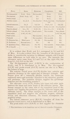
Volume 1
On the anatomy, physiology, and pathology of the chimpanzee / by Charles F. Sonntag.
- Sonntag, Charles F. (Charles Frederick), -1925
- Date:
- 1923
Licence: In copyright
Credit: On the anatomy, physiology, and pathology of the chimpanzee / by Charles F. Sonntag. Source: Wellcome Collection.
96/118 page 416
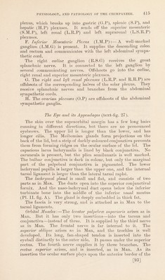
![superior oblique. It passes through an arch formed by the capsule of Tenon. The rectus externus arises by two heads and is inserted as in Man. It is broad and moderately thick. The third nerve crosses both heads instead of passing between them. The fourth nerve passes over both heads as in Man. And the sixth nerve comes out between them before sinking into their ocular surface. The naso-ciliary nerve also crosses both heads. The rectus interims is broad and thick, and its attachments are as in Man. Its nerve, from the superior division of the oculomotor nerve supplies it by several twigs. The inferior oblique arises by fleshy and tendinous fibres from the floor of the orbit a quarter of an inch external to the naso- lachrymal duct. It is not spread out as in Man, but remains as a thin belly, which is inserted farther back into the sclera close to the entrance of the optic nerve on the postero-lateral aspect of the ball (text-fig. 27). The rectus inferior is as in Man. It is, therefore evident that the recti are almost as in Man, but the obliques and levator palpebraB differ. The nerves and vessels are described in other sections of this paper. The capsule of Tenon is very strong. The ophthalmic veins are as in Man. On pulling the eye forwards it was seen that the fascia lying- next to the eyeball was seen to be well developed, and almost free from fat. The globe itself is relatively smaller than in Man, but the ophthalmoscopic appearances are very similar in both, as pointed out by Lindsay Johnstone (70). Auditory Apparatus. It is well known that the auricle is less degenerate in the Chimpanzee than in Man and the other Anthropoids. And from the numerous accounts which have been published it appears that the auricle is one of the most variable parts of the external anatomy of the Chimpanzee. Its very complete form in my specimen is shown in Plate I. fig. A. It has few hairs, and Wallis (58) pointed out that it has this feature in all examples. Darwin (16) noted that neither the Orang nor the Chimpanzee move their auricles, and I was unable to detect any movements on any occasion when I made observations in the Ape House in the Gardens. In Plate II. fig. B it is shown how the auricular cartilage is very complete, and it has a wide, thin peripheral rim. But the human auricular cartilage is a totally different thing. I was unable to detect intrinsic muscles in the cartilage. The tympanic membrane cannot be seen through the ordinary aural specula, for it lies at the end of a long, bony external auditory meatus. The Eustachian tnbe has no well-marked torus round its pharyngeal end, and I did not detect a salpingo-pharyngeus muscle. [94]](https://iiif.wellcomecollection.org/image/b2982123x_0001_0096.jp2/full/800%2C/0/default.jpg)
 No text description is available for this image
 No text description is available for this image
 No text description is available for this image