A manual of operative surgery / By Lewis A. Stimson ... and John Rogers.
- Lewis Atterbury Stimson
- Date:
- 1900
Licence: Public Domain Mark
Credit: A manual of operative surgery / By Lewis A. Stimson ... and John Rogers. Source: Wellcome Collection.
Provider: This material has been provided by the Augustus C. Long Health Sciences Library at Columbia University and Columbia University Libraries/Information Services, through the Medical Heritage Library. The original may be consulted at the the Augustus C. Long Health Sciences Library at Columbia University and Columbia University.
143/628 (page 139)
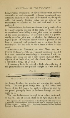
![Fig. 50. and then behind that bone, and carried upward so as to divide the upper capsular attachments in front and behind. 4. A pair of bone-forceps are next employed to cut off the entire inner condyle and trochlea of the humerus [from above downward], and then introduced in the opposite direction [from below upward and outward], so as to de- tach the external condyle and capitellum of the humerus from the shaft. 5. The angular end of the humerus is turned out through the incision and sawn off' square, 6. The external condyle and capitellum are removed partly by twisting, partly by dissection, without any division of the skin on the outer side of the arm. If there is dense osseous union that cannot be overcome by flexion and extension under chloroform, the humerus must be divided through the condyle with bone- pliers, and the operation completed as above. Operative Reduction of Old Unreduced Backward Dislo- cation of the Elbow.1—The first incision is made on the outer side (Fig. 50), beginning well up (in the supinator ridge and passing downward to and across the head of the radius, and then for one or two inches posteriorly in the interval between the radius and ulna. Through this the newly formed bone (Fig. 50, A) on the back of the humerus is exposed and chiseled away, and the outer aspect of the external con- dyle freed by dividing its fibrous attachments to the radius and ulna until the capitellum is freely exposed. The sides of the upper portion of the wound are then retracted, the olecranon exposed, and the sigmoid cavity cleared of the mass of fibrous tissue which, more or less, fills it and binds it to the back of the humerus. »L. A. Stimson: N. Y. Med. Journ., Oct. 24, 1891. Incision for the operative treatment of old unreduced dis- location of the elbow. A. Peri- osteal bridge and new tissue occupying the posterior surface of the lower extremity of the humerus.](https://iiif.wellcomecollection.org/image/b2120651x_0143.jp2/full/800%2C/0/default.jpg)