The practice of medicine and surgery : applied to the diseases and accidents incident to women / by W. H. Byford ... and Henry T. Byford.
- William Heath Byford
- Date:
- 1888
Licence: Public Domain Mark
Credit: The practice of medicine and surgery : applied to the diseases and accidents incident to women / by W. H. Byford ... and Henry T. Byford. Source: Wellcome Collection.
Provider: This material has been provided by the Francis A. Countway Library of Medicine, through the Medical Heritage Library. The original may be consulted at the Francis A. Countway Library of Medicine, Harvard Medical School.
42/876 (page 26)
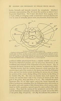
![fascia3, forwards and inwards towards the symphysis. Schultze's schematic representation (Fig. 10) shows the position in which I have frequently found tliem. Yet I have also found them sagging down or back a little, or swinging a trifle around their external attachments even in cases of normally placed uteri, and, therefore, think that their Fig. 10. Position of the Ovaries (after Schultze) {]/Q. a, fundus of uterus behind pubes ; b, fundus wtien the bladder is full; c, sacrum ; ap, anterior superior spine of ileum ; ps, edge of psoas muscle ; ip, infundibulo-pelvic ligament; ov^, nor- mally placed OYary ; ov, ovary drawn back into the hollow of the sacrum by displaced fundus uteri ; ov^, ovary drawn back beside cervix by the replaced fundus ; ov*, ovary pressed or held forward when fundus is back; ft, Fallopian tube; ol, ovarian ligament. position is within physiological limits a slightly variable one, and is affected by abdominal pressure, and by temporary alterations in the conditions and relations of the abdominal and pelvic viscera. Fig. 11 represents the broad ligament and its contents, modified from Henle. Schultze teaches that the ovarian ligaments, which pass from the anterior-inner end of the ovaries to the uterus just below the Fallopian tubes, and are four inches across (including the uterus), do not change the position of the ovaries during lifting of the fundus by the filling of the bladder (Fig. 10). But when the fundus leans back against the sacrum, the anterior inner ends of the ovaries are drawn to the back part of the pelvis ; they pass from ov^ to ov\ The infundibulo-pelvic ligaments (Fig. 11) or outer upper end of the broad ligaments are folds of peritoneum extending from the Fallopian tubes and ovaries to the pelvic wall, and contain a little fibrous tissue, which passes, some- times in visible quantities, upward upon the outer surface of the peri- toneum. They limit the motion of the peripheral end of the ovary and the fiml^riated extremity of the tubes to a small area at the sides of the pelvis (Figs. 10 and 12). Ov^, Fig. 10, indicates the position of](https://iiif.wellcomecollection.org/image/b21044958_0042.jp2/full/800%2C/0/default.jpg)