Volume 1
Operative gynecology / by Howard A. Kelly.
- Howard Atwood Kelly
- Date:
- 1901, ©1898
Licence: Attribution-NonCommercial 4.0 International (CC BY-NC 4.0)
Credit: Operative gynecology / by Howard A. Kelly. Source: Wellcome Collection.
Provider: This material has been provided by The University of Glasgow Library. The original may be consulted at The University of Glasgow Library.
533/622 (page 489)
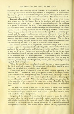
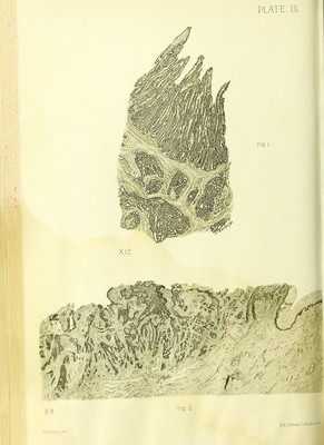
![TUBERCULOSIS OF THE ENDOMETRIUM. 489 packing its cavity loosely with gauze, tlie ends of which are allowed to hang out of the cervix into the vagina; my own practice, however, is simply to place a loose gauze pack in the vagina, which is renewed every twenty-four hours. Patients should be kept in bed after curetting for abortion for two weeks or longer, to allow involution of the uterus to take place; care of the patient is just as important at this time as in the puerperiuui after a normal labor. Microscopic Examination for the Remnants of an Abor- tion.—We usually have in these cases the clinical history of a recent miscar- riage, and the amount of material removed by curettage is often abundant. As a rule, there is no suggestion as to their source in the macroscopic appearance of the tissues; occasionally little villous threads can be seen. Histologically, the appearance of glandular hypertrophy predominates; the glands are dilated, convoluted, and show little titlike processes springing into their lumina; the epithelium is a little flattened and the stroma of the mucosa shows marked swelling of its cells in the superficial portion, forming typical decidual cells which persist for several weeks after the abortion. These appearances are suggestive of pregnancy, but a positive diagnosis must rest upon the discovery of villi; these in the early months still show two layers of epithelial covering, the inner of which is made up of cuboidal cells; the outer syncytial layer appears as a ribbon of protoplasm with nuclei distributed through it; this outer layer sends out protoplasmic buds which form the new villi, and in the centers of these buds are found from five to forty nuclei, forming the so- called placental giant cells, because when cut across they present the appearance of a typical giant cell. The interior of a villus is composed of mucoid tissue rich in blood vessels. In one obscure case nothing was found in the curettings but some glandular hypertrophy, ill-defined decidual cells, and a single free giant cell; this latter structure led to a further searching investigation, which was rewarded by the discovery of villi, confirming the diagnosis of pregnancy. Tuberculosis of the Endometrium.—In the early stages the epithelium of the surface is intact, the glands normal, and the tubercles are found scattered throughout the superficial portions of the stroma, consisting of aggregations of epithelioid cells; later they are surrounded by small round cells, and at a still later date ffiant cells are found in the center. The surface epithelium over a superficial nodule is often somewhat flattened and pale. In a marked case the glands are encroached upon, and it is at times almost impossible to distinguish some of the epithelioid cells from the gland epi- thelium ; in other glands, tubercles are seen partly projecting into and obliter- ating the cavity; again the gland may be filled with caseous material. In the most advanced cases where the cavity of flip uterus is lined by caseous material, the surface is covered by a necrotiakmateriai devoid of nuclei, below which lies a zon^ of typical tuberculous tissu^consisting of epithelioid cells and tubercles ; in the deeper portions a stray gland may survive; where the process has gone deep enough tdftivolve the musc]e,^e glands are often entirely absent. Bacilli are found \mh varying frequency, sometimes sparse, sometimes 35 1](https://iiif.wellcomecollection.org/image/b21466099_0001_0547.jp2/full/800%2C/0/default.jpg)