Hunterian lectures on the morbid anatomy, pathology, and treatment of hernia / by Charles Barrett Lockwood.
- Date:
- 1889
Licence: Public Domain Mark
Credit: Hunterian lectures on the morbid anatomy, pathology, and treatment of hernia / by Charles Barrett Lockwood. Source: Wellcome Collection.
Provider: This material has been provided by the Royal College of Physicians of Edinburgh. The original may be consulted at the Royal College of Physicians of Edinburgh.
145/216 (page 133)
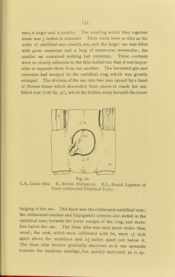
![]33 sacs, a larger and a smaller. The swelling which they together made was 3 inches in diameter. Their walls were as thin as the walls of umbilical sacs usually are, and the larger sac was filled with great omentum and a loop of transverse mesocolon; the smaller sac contained nothing but omentum. These contents were so closely adherent to the thin-walled sac that it was impos- sible to separate them from one another. The herniated gut and omentum had escaped by the umbilical ring, which was greatly enlarged. The division of the sac into two was caused by a band of fibrous tissue which descended from above to reach the um- bilical scar {vide fig. 30), which lay hidden away beneath the lower Fig. 30. L.A., Linea Alba. R., Rectus Abdominis. R.L., Round Ligment of Liver (obliterated Umbilical Vein). bulging of the sac. This band was the obliterated umbilical vein ; the obliterated urachus and hypogastric arteries also ended in the umbilical scar, towards the lower margin of the ring, and there- fore below the sac. The linea alba was very much wider than usual; the recti, which were infiltrated with fat, were i| inch apart above the umbilicus and z\ inches apart just below it. The linea alba became gradually narrower as it ran upwards towards the ensiform cartilage, but quickly narrowed as it ap-](https://iiif.wellcomecollection.org/image/b21911897_0145.jp2/full/800%2C/0/default.jpg)