Text-book of ophthalmology / by Dr. Ernest Fuchs ... authorized translation, revised from the seventh enlarged and improved German edition by A. Duane ... With two hundred and seventy-seven illustrations.
- Ernst Fuchs
- Date:
- 1899
Licence: Public Domain Mark
Credit: Text-book of ophthalmology / by Dr. Ernest Fuchs ... authorized translation, revised from the seventh enlarged and improved German edition by A. Duane ... With two hundred and seventy-seven illustrations. Source: Wellcome Collection.
Provider: This material has been provided by the Harvey Cushing/John Hay Whitney Medical Library at Yale University, through the Medical Heritage Library. The original may be consulted at the Harvey Cushing/John Hay Whitney Medical Library at Yale University.
43/892 page 21
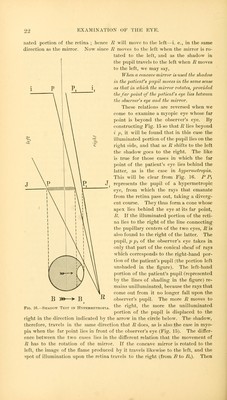
![2] the left of this Hue. From R the rays (now become divergent) continue on their way toward the observer's eye, which we will now suppose to be beyond B. / p and p\ ii represent the iris, and p p, the pupil of this eye. Now p px does not take in all of the conical sheaf of rays emanating from R, but only a por- tion of it, having p o as its base. The remainder of the cone falls upon the iris p i. Since the rays constituting this part of the cone are not seen by the observer, the portion of the pupil which is opposite to them, and from which they come (represented in Fig. 15 by the lines of shading), appears unillu- minated ; the only portion of the pupil that does appear illuminated being that which is here shown as unshaded, and from which the observer receives rays that enter his own pupil. The dark and the luminous portions of the pa- tient's pupil are separated by a curved line, since the boundary between the two is formed by the pupillary edge p of the observer's eye. Thus the circle at the bottom of Fig. 15 represents the pupil of the patient's eye seen from in front ; the portion of it left unshaded in the figure corresponds to the illumi- nated part of the pupil. Now suppose that by a rotation of the mirror the spot of illumination in the fundus shifts in such a way that R travels farther to the left. Then more and more of the emergent beam will fall upon the iris, and less and less of it will fall upon the pupil of the ob- server's eye, and the shadow in the pupil of the patient's eye will, as the arrow in the circle indicates, advance farther and farther toward the left pu- pillary margin, until finally the whole pupil appears dark. The shadow, therefore, moves in the same direction that R does. We have now to determine how the movements of R are related to the movements of the mirror. If a concave mirror is employed, it forms at its focus an image of the lamp flame which lies between the mirror and the pa- tient's eye and serves to illuminate the latter. If the mirror is rotated to the left, the image of the flame also travels to the left. But as the portion of retina illuminated must always lie on the side diametrically opposite to the body that illuminates it—namely, the image of the flame—it must, with the movements of the mirror, move in a sense opposed to that of the image of the flame—i. e., to the right (from B or Pi in Fig. 15). But the point of union, R, of the emergent rays lies diametrically opposite to that occupied by the illumi- B>»-->B](https://iiif.wellcomecollection.org/image/b21023906_0043.jp2/full/800%2C/0/default.jpg)


 No text description is available for this image
 No text description is available for this image
 No text description is available for this image