The Anatomy and physiology of the organ of hearing : with remarks on congenital deafness, the diseases of the ear, some imperfections of the organ of speech, and the proper treatment of these several affections / By David Tod.
- Tod, David
- Date:
- 1832
Licence: Public Domain Mark
Credit: The Anatomy and physiology of the organ of hearing : with remarks on congenital deafness, the diseases of the ear, some imperfections of the organ of speech, and the proper treatment of these several affections / By David Tod. Source: Wellcome Collection.
Provider: This material has been provided by The University of Leeds Library. The original may be consulted at The University of Leeds Library.
15/174
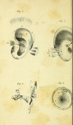
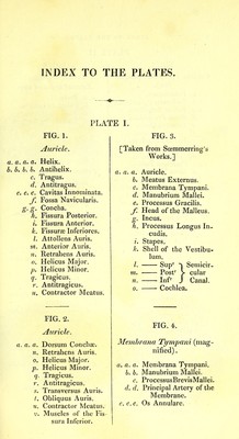
![INDEX TO THE PLATES. PLATE I. FIG. 1. Auricle. a. a. a, a. Helix. b. b, b. b. Antihelix. c. Tragus. d. Antitragus. e. e, e. Cavitas Innominata. Fossa Navicularis. g.g. Concha. h. Fissura Posterior. i. Fissura Anterior. k. Fissurse Inferiores. I. Attollens Auris. m. Anterior Auris. n. Retrahens Auris. 0. Helicus Major. p. Helicus Minor. q. Tragicus. r. Antitragicus. u. Contractor Meatus. FIG. 2. Auricle. a, a. n. 0. V- r. Dorsum ConchiE. Retrahens Auris. Helicus Major. Helicus Minor. Tragicus. Antitragicus. s, Transversus Auris, /. Obliquus Auris. u. Contractor Meatus. V. Muscles of the Fis- sura Inferior. FIG. 3. [Taken from Sosmmerring's Works.] a. a. a. Auricle. b. Meatus Externus. c. Membrana Tympani, d. Manubrium Mallei. e. Processus Gracilis. /■ Head of the Malleus. Incus. f. Processus Longus In- cudis. i. Stapes. k. Shell of the Vestibu- lum. I. —— Sup^ ^ Semicir- m. Post^ > cular n. Inf^ J Canal. 0. Cochlea. FIG. 4. 3Iembrana Tympani (mag- nified) . a, a. a. Membrana Tympani. 6. b. Manubrium Mallei. c. ProcessusBrevisMallei, d. d. Principal Artery of the Membrane. e. e. e. Os Annulare.](https://iiif.wellcomecollection.org/image/b21514203_0015.jp2/full/800%2C/0/default.jpg)