Licence: Public Domain Mark
Credit: A text book of physiology / by William Rutherford. Source: Wellcome Collection.
Provider: This material has been provided by the Royal College of Physicians of Edinburgh. The original may be consulted at the Royal College of Physicians of Edinburgh.
112/180 (page 100)
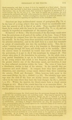
![osteoblasts. As age advances the inner layer of the periosteum becomes more fibrous, and the osteoblasts fewer in number (Fig. 73). White and elastic fibres pass from the periosteum into the bone, and constitute the perforating fibres. The periosteum is an exceed- ingly vascular membrane, most of its blood-vessels being destined for the bone. It also contains numerous lymphatics and nerves, many of which accompany the blood-vessels into the bone. The deeper layer of the periosteum is prolonged Fig. 73. 2), Inner layer of • , ,1 -rj • i t i.- 1 ? periosteum of femur of mto ail the Haversian canals. In a vertical section adult eat; 6, osseous tissue, of one of these ill a softened bone, the vessels, con- nective tissue, and osteoblasts, may be readily seen if the bone be young (Fig. 72, d); but in adult bone the osteoblasts are few in number. A Haversian canal therefore contains blood-vessels, fine con- nective tissue, osteoblasts, together M'ith lymphatics and nerves. On the inner aspect of the bone the canals open into the medullary spaces of the spongy bone, and the osteoblasts and vascular connective tissue are pro- longed through the canals into the cancelli. Everywhere in the interior of the cancelli of spongy bone, and the medullary canal of a long bone, there is a layer of cells, mostly osteoblasts (Fig. 72, g), but in spongy bone, particularly in that which is young, these are here and there replaced by large multinucleated giant cells, which, according to Kolliker, are con- cerned, not in the formation, but in the absorption of bone, hence he has named them osteoclasts (Fig. 79, oc). In the medullary canal of a long bone the vascular fibrous tissue in contact with the bone forms a sort of membrane, that has received the special name of endosteum. The ]\Tai?row of bono is either yellow or red. Yellow manoio occurs in the medullary canal, and larger cancelli of long bones. It chiefly con- sists of fat-cells and blood-vessels, with some ^ connective tissue. Red marroio occurs in the |' ~ spongy tissue at the ends of the long bones of /'■ the limbs, in the clavicle, ribs, and in all the flat j and short bones. It consists-of a delicate con- | \ nective tissue, with many blood-vessels and a ? great number of cells, most of which are the medullary cells or proper marrow cells of Kol- liker. They are all nucleated i)rotoplasts, many of them exactly resembling lymph-corpuscles (Fig. 74, g, d), while others have a single large nucleus, with a nucleolus (c). Others closely resemble these, but contain fatty particles, and appear to be young fat cells (.); red marrow, ^J^^J^^^^ZH however, contains very few fat cells, ihere are q-^^^^ ^^^is h, coiouied biood- manv red blood-corpuscles (b), but they probably corpuscle; c, d, e, /, g meciuiiaiT , , , 11 1 -^1 • \-i 1 „■(• cells. X 300. (The nuclei of the cells belong to the blood withm the vessels of the l^^^ ^^^J^^ ^^^^^^ marrow. aicohoi.) Neumann and Bizzozero {Op. 23, year 1868, pp. 689, 885) state that in I'ed marrow tliere are nucleated red biood-corpuscles similar to tliose found in tlie embryo (J? ig.^ J a;. From tliis tliey conclude that in red marrow, white corpuscles develop mto coiomea](https://iiif.wellcomecollection.org/image/b21981747_0112.jp2/full/800%2C/0/default.jpg)