Licence: Public Domain Mark
Credit: A text book of physiology / by William Rutherford. Source: Wellcome Collection.
Provider: This material has been provided by the Royal College of Physicians of Edinburgh. The original may be consulted at the Royal College of Physicians of Edinburgh.
80/180 (page 68)
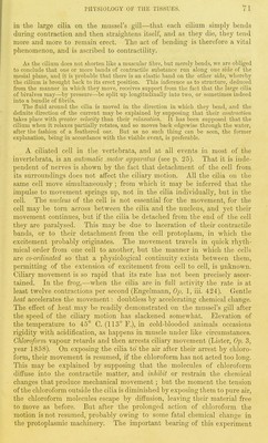
![are due to a reduction of the silver salt in what appears to be an inter- stitial or cementing substance. Prolonged action of the silver solution, however, is followed by darkening of the general cell-substance as well as of the cement. We owe to His and ]iecklinghausen this method which has already proved of great value in the study of blood-capillaries and the lymphatic system. Endothelial plates are probably all nucleated at an early period of development, and the nucleus is often per- manent, but it disappears from many cells as they grow old (Fig. 29, c). The duration of endothelial scales and the mode in which they are replaced are points of much obscurity. In the air-ve.sicles small nucleated finely- granular cells (Fig. 29, b), apparently of a germinal character, may be seen at intervals amidst the large clear plates, and thougli division of the small cells may sometimes be seen {b), it is doubtful whether or not they expand and replace the large ones. In blood-vessels, however, these minute germinal cells are wanting, and nothing can be seen but a layer of clear plates similar to the larger scales in the air vesicles, but mostly nucleated. Fig. 29. Silvered ciidotheliuin of vesicles of lung of cat. a. Outline of air-vesielo ; c, old endothelial scale ; ft, nucleated llnoly-grnnulnr cells. X 300. COLUMNAR EPITHELIUM. Columnar epithelium lines the alimentary canal below the oesophagus, and the greater part of the ducts opening into the canal, including Fig. 30. Columnar epithelium from small intestine of cat. a, Striated border at free end of cell, ft. Chalice cell, x 600. Fig. 31. Fresh epi- thelium from cat's large intestine, after magenta staining. a. Ordinary columnar cell with border split up by artificial pressure. 6, Chalice cell. X 600. (The two cells are in exact relative pro- portion.) Fig. 32. Surface view of free ends of columnar epithelium from villas of frog's intestine, silvered, a. Opening of chalice cell, ft. Periphery of the broader subjacent part of the cell. X 400. the gall-bladder. It is also found in the olfactory region of the nose, in the urethra, and vas deferens. It exists everywhere as a single layer,](https://iiif.wellcomecollection.org/image/b21981747_0080.jp2/full/800%2C/0/default.jpg)