Licence: Public Domain Mark
Credit: A text book of physiology / by William Rutherford. Source: Wellcome Collection.
Provider: This material has been provided by the Royal College of Physicians of Edinburgh. The original may be consulted at the Royal College of Physicians of Edinburgh.
88/180 (page 76)
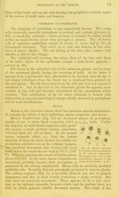
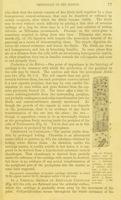
![TEXT-BOOK OF PHYSIOLOGY. solution of alum (1 : 200), shows that the tissue consists of nucleated cells imbedded in a hyaline matrix (Fig. 39). The Cells are more or less rounded, and often somewhat anguhir. Each cell (a) is a mass of finely-granular protoplasm with a well-defined ,.^v:■^'r''':r:;v^sM'^■'■'■■^'^'.■■;^.,:,,,.., spherical nucleus, and often two or or three nucleoli. The protoplasm entirely fills the cell space, but if water and various other re-agents be added, it shrinks (e), and, gathering around the nucleus, leaves a clear space in the periphery of the cell cavity: proving thai 2yi'otoplasm is a sponge-like substance \vith inter- stices full of fluid. When the pro- toplasm shrinks from the capsule there is no evidence of a laceration of the protoplasm, as must have occurred, were the capsule a UK^re hardening or other transformation of the outer part of the protoplasm, The cells as some have imagined. Pig. 39. Aii,ii;iilar cjulila-f liMiii lioail uf frog femur. Examined in alum solution (1 : 200). a. Typi- cal cell with clear cniisule ; b, cell with divided nucleu.s ; c, cell with divided nucleus and proto- ^ . ^ plasm; (/, two colls within a primary capsule, each lH'iy be StaiUcd by VariOUS ageuts, 01 with a secondary capsule ; P, cell with protoplasm which qold-clllorkU is pPrhapS the shrivelled around the nucleus, x 800. (The proto- plasm in e is too faint. It ought to have been drawn darker than in the other cell.4, somewhat liko b and c in I''ig. 21.) best. It dyes the cells violet, with- out causing them to shrink. lod'me solution stains the cell substance pale yellow, and reveals the presence of ^;(tr//c/«s of glycogen by rendering them brown. Osniic acid slightly darkens the general cell substance, and renders hlack any fatty particles that may be present. The trniismission of electrical shocks througli a thin ]jlato of living cartilage, e.g. the eu.sifonn cartilage of the newt, occasions a sudden .shrinking of the cells, such as happens when a white blood-corpuscle is thus excited. B)' Rollctt (Op. 38, i. p. 98) the shrinking is ascribed to contraction ; but Hcidonbain [Op. 49, Heft 2, p. 1), who discovered this fact, maintains that as the protopla.sm never relaxes and refills the cell-space, the electricity, like water and other re-agents, simply occasions coagulation of the pi'oteids in the protoplasm. The Matrix {intercellnlar substance or p)eriplast) has the appearance of ground glass. It is hj^aline, faintly granular, and Avithout special methods of preparation appears homogeneous. It is secreted by the cells, around each one of which the newest part of the matrix constitutes a clear homogeneous capsule, at first distinct, but becoming after a time so fused with the surrounding matrix as to be undistinguishable from it. The matrix may be stained of a brown colour by the silver process, and by this means the capsule around each cell is often very clearly defined. Outside the cell-capsule the matrix appears homogeneous, but is in reality fibrillated, as was first pointed out by Leidy, an American histologist. The fibrillation may become very evident in some pathological states of cartilage, as Eedfern was the first to show {Op. 7, August 1849). Eecently this point has been reinvestigated by Tillmanns {Op. 18, x. p. 401; {Op. 16, Anat. Abtheil., year 1877, p. 9) and Baber {Op. 1, x. p. 113).](https://iiif.wellcomecollection.org/image/b21981747_0088.jp2/full/800%2C/0/default.jpg)