Licence: Public Domain Mark
Credit: A text book of physiology / by William Rutherford. Source: Wellcome Collection.
Provider: This material has been provided by the Royal College of Physicians of Edinburgh. The original may be consulted at the Royal College of Physicians of Edinburgh.
99/180 (page 87)
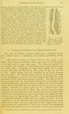
![with tlie other cells of connective tissue. They (Fig. 51, d) consist of a nucleated mass of coai'.sely-grauular protoplasm, three or four times the size of the wandering cells; usually, however, there is no amoeboid movement; but in the delicate intermuscular fascia of the frog's leg they may be seen to slowly change their form (Kiihue). They have no distinct piocesses, and are usually more or less rounded in shape. In ordinary areolar tissue they may be found in small number, usually in gioups around the blood-vessels. Fat sometimes appears within them, and they become trans- formed into fat cells. Waldeyer (0^. 18, xi. p. 190) proposes to designate these cells the embryonal cells of connective tissue, or plasma cells; but both terms are objectionable, because, as he himself points out, they may be,developed from oi'dinary connective tissue cells: and certainly the term plasma is too ambiguous. Boll {Op. 18, vii. p. 322) pointed out the similarity between these cells and the so-called interstitial cells of the testis; and Waldeyer cit.) refers to the same category other peculiar groups of cells, whose histological significance has hitherto been obscure — viz. the cells of the coccygeal body (Fig. 5i), corpus luteum, suprarenal capsule, and the serotinal ^.j^^ cells of the placenta. Waldeyer states that in all these situa- '^'^ .' i ^ capil- tions the cells are developed from ordinary connective tissue „ f,-,,! ii'„o,,„ 1 11 1 ii 1 1 1 1 /T71- mi lary; fi, connective tissue corpuscles, and always envelope the blood-vessels (Fig. 54). The g,,4tl. • h coarsely-gian- fuuctions, if any, of the cells in these organs are unlcuowu. ^.^^^ ^^[-^ ' (E|,erUi) 2. Arrangement of the Elements of ordinary Connective Tissue. The fibres of ordinary connective tissue may be arranged loosely, as in areolar tissue; or compactly, as in tendon, aponeurosis, cornea, etc. a. Thk Areolar Form of Fibrous Tissue.—The areolar is the most common form of connective tissue, and occurs in the deep layer of the .skin, and in many other parts. The fibres are arranged in an irregular open network, the white being much more numerous than the elastic fibres. Sometimes an elastic fibre appears to lie within a white fibre (Key and Ketzius, Op. 52, i. plate 14, fig. 12), but this is rare. Almost always they run irregularly in the spaces between the white fibres. The irregular spaces between the fibres are filled with lymph (lymph spaces), and by a free intercommunication allow of a ready percolation of the lymph from one part to another. The effect of this is seen in dropsy, wliere the lymph drains through the areolar tissue to the most dependent part; and also in cutaneous emphysema, when the lymph spaces become distended with air, which freely moves from one part to another. Normally, the lymph spaces are in a collapsed state. The amoeboid cells wander about in them. The ordinary connective tissue corpuscles ai-e also within them, but not lying loose as they appear after dislocation by artificial means, but in contact with and dasimig the white fibres as shown by Eanvier {Op. 39, p. 342). They may clasp and partly or completely surround a single fibre, or a bundle of fibres. In some situations, e.g. in the omentum and in the subarachnoid space, they are epithelioid in shape, and by the apposition of their margins form a com- plete envelope to a single fibre, as seen in the tissue of the subarachnoid space (Key and Retzius, Op. cit), or to bundles of fibres, as occurs in the ^ A description of the coccygeal body by Eberth may be found in 0]). 38, i. p. 295.](https://iiif.wellcomecollection.org/image/b21981747_0099.jp2/full/800%2C/0/default.jpg)