Observations on certain parts of the animal œconomy / by John Hunter.
- John Hunter
- Date:
- 1786
Licence: Public Domain Mark
Credit: Observations on certain parts of the animal œconomy / by John Hunter. Source: Wellcome Collection.
Provider: This material has been provided by The University of Leeds Library. The original may be consulted at The University of Leeds Library.
43/490 (page 23)
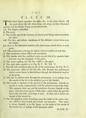
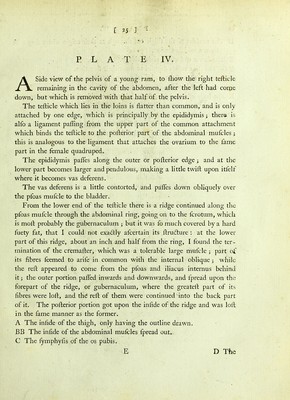
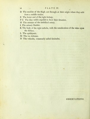
![[ 23 ] PLATE III. THE third figure reprefents the teftes, &c. in the fame fubjedt; all the parts above the ofla ilium being cut away, and the abdominal mufcles and the bladder being turned downwards. AA The thighs, unfiniftied. B The penis. C The middle part of the fcrotum, its lateral parts being removed to fhow the teftes. DD The fkin and cellular membrane of the abdomen turned down over the thighs. EE Part of the abdominal mufcles and peritonaeum turned down at each groin. FF The peritonaeum covering the iliacus internus mufcle of each fide. G The inteftinum redtum filled with meconium. H The bladder with the umbilical artery on each fide of it, turned a little forwards over the fymphyfis of the pubes. II The ureters pafling over the iliac veflels to the pelvis. K The right teftis expofed, as in Fig. II. V. W. XX. Y. L The left teftis inclofed in the procefs of the peritonaeum. See Fig. II. U. M The Tpermatic veflelsv: of the feft fide, feen through the peritonaeum which covers them, in their, defcent through the abdominal mufcles at the groin. N The left vas deferens feen through the peritonaeum, in its paflage from the mouth of the fac to the pofterior part of the bladder. O The mouth or aperture of the procefs of the peritonaeum, whereby its mouth or cavity communicates with the general cavity of the belly. This aperture clofes up, and the membrane becomes fmooth at this place, when the foetus grows a little older unlefs when the gut falls down after the teftis, and keeps it open. In that cafe it makes the mouth of the hernial fac. P The left epigaftric artery branching upon the infide of the redtus muf- cle, which is here turned downwards and outwards. This artery is always fituated, as in this figure, on the infide of the mouth of the hernial fac, or paflage of the fpermatic veflels. PLATE IV.](https://iiif.wellcomecollection.org/image/b2151673x_0043.jp2/full/800%2C/0/default.jpg)