Variations in human myology observed during the winter session of 1867-68 at King's College, London / by John Wood.
- John Wood
- Date:
- 1868
Licence: Public Domain Mark
Credit: Variations in human myology observed during the winter session of 1867-68 at King's College, London / by John Wood. Source: Wellcome Collection.
Provider: This material has been provided by The Royal College of Surgeons of England. The original may be consulted at The Royal College of Surgeons of England.
36/46 page 516
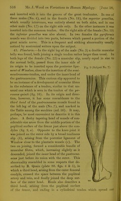
![and inserted with it into the groove of the great trochanter. In one of these males (No. 4), and in the female (No. 19), the superior gemellus, which usually intervenes, was entirely absent on both sides, and in one other male (No. 17) on the right side only. In the other instances it was inserted into the common tendon. On the right side of the female (No. 19) the inferior gemellus was also absent. In two females the pyriformis muscle was divided into two parts, between which passed a portion of the great sciatic nerve. This is a frequent and striking abnormality usually noticed by anatomical writers upon the subject, 43. Planlaris.—In the right leg of the male (No. 3) a double muscular belly was found, both joining a single tendon rather larger than usual. In both legs of the female (No. 22) a muscular slip, nearly equal in size to the normal belly, passed from the inner side of its origin to be inserted upon the ^os^erio?'/«]^a- Fig. 9 (Subject No. 7). ment of Winslow, close to the insertion of the semi- inetnbranosus-tendon, and under the inner head of the gastrocnemius. This curious slip appeared to be an instance of a development of muscular fibres in the substance of a tendon, similar to that un- usual one which is seen in the tendon of the pe- roneiis quinti (fig. 10). In its origin and direc- tion, however, it has some resemblance to the third head of the gastrocnemius muscle found in the left leg of the male (No. 7), and marked in the Table among the sundries (col. 56). It may, perhaps, be most convenient to describe it in this place. A fleshy tapering head of muscle of con- siderable size arose from the middle portion of the popliteal surface of the femur just above the con- dyles (fig. 9, a). Opposite to the knee-joint it was joined on the outer side by a broad tendinous slip (6), arising from the posterior ligament of Winslow close to the plantaris muscle (c). The two on joining, formed a considerable bundle of muscular fibres, which, increasing slightly as it descended, joined the inner head of the gastrocne- mius just before its union with the outer. This abnormality resembled in some respects that de- scribed by R. Quain (plate 80. figs. 4 & 5), in which a third head, arising from the outer femoral condyle, crossed the space between the popliteal artery and vein, and finally joined the deep sur- face of the outer head. Ilenle also describes a third head, arising from the popliteal surface of the femur, and ending in a cylindrical tendon which spread out](https://iiif.wellcomecollection.org/image/b2227375x_0036.jp2/full/800%2C/0/default.jpg)