The house fly : Musca domestica, Linnæus a study of its structure, development, bionomics and economy / by C. Gordon Hewitt.
- Hewitt, C. Gordon (Charles Gordon), 1885-1920.
- Date:
- 1910
Licence: In copyright
Credit: The house fly : Musca domestica, Linnæus a study of its structure, development, bionomics and economy / by C. Gordon Hewitt. Source: Wellcome Collection.
224/256 (page 173)
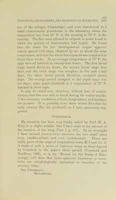
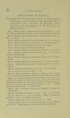
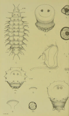
![414 (\ flOKDON IIF.WI'IT, KXPLANA'riON OF PT.A'I'E 22, Illustrating Dr. C. Oorclon Hewitt’s paper on “The Structure, Development, ami Bionomics of the Ilon.se-fly, Mu.sca domestica, Linn. Part TIT. The Bionomics, Allies, Parasites, and the Delations of ^f. domestica lo Human Disease.” Fig.].—Matai’e larva of Homalomyia canicnlaris, L. x 17. a.sp. Anterior spiracnlar processes. p..sp. Postenorspii-acular aj;ei-tnres. Fig. 2.— Posterior end of mature lami of Anthomyia radicnm Mg. an. Anns. Fig. 3.—Anterior spiracnlar process of mature larva of A. radicnm. Fig. 4.—Head of Stomoxys calcitrans, L.; left lateral a.spect. Fig. 5.—Postei’ior end of matime laiva of S. calcitrans. Fig. 6.—Posterior spiracle of the same, enlarged. Fig. 7.—Posterior spiracle of mature larva of Musca domestica. Fig. 8.—Posterior spiracles of first larval stage of Callipliora erythrocepliala, Mg. Fig. 9.—Posterior s^jiracles of second larval stage of C. erythro- cephala. Fig. 10.—Posterior spiracle of mature larva of C. ery throcepliala. Fig. 11.—Anterior spiracnlar process of mature larva of C. erythro- cephala. Fig. 12.—Posterior end of mature larva of C. erythrocephala. Fi. 13.—Chernes nodosus. Schr. x 30. Fig. 14.—Thoraco-ahdominal region of Homalomyia canicu- lar is, ? . showing Gamasids attached to the ventral side of the ahdomcn. Fig. 15.—Longitudinal (sagittal) section of abdomen of M. dom es t i ca. which has been killed by Empusa musca;, showing the feltwork of fungal hypha; filling the inside of the abdominal cavity and the pro- duction of conidia in the intersegmental regions. X 12. c. Conidio- pliores producing conidia. /. Fungal hypha'. Fig. 10.—Four conidiophores showing the formation of conidia (r.). X 100 (approx.). Fig. 17. —Conidium of Empusa musca;. X 400. o.<j. Oil globule. Pig 18.—Habronema musca; (Carter). Adult but iminatun? specimen. X 85. <j.a. Genito-anal aportin-e. Fi^r, 19.—Caudal end of Habronema musca;. X 300. Fig. 20.—Tarsal Joints of one of posterior pair of legs of Musca domestica. Lateral aspect, to show densely setaceous character.](https://iiif.wellcomecollection.org/image/b28047436_0224.jp2/full/800%2C/0/default.jpg)