Licence: Public Domain Mark
Credit: A practical guide to meat inspection / by Thomas Walley. Source: Wellcome Collection.
Provider: This material has been provided by the Royal College of Physicians of Edinburgh. The original may be consulted at the Royal College of Physicians of Edinburgh.
39/230 (page 27)
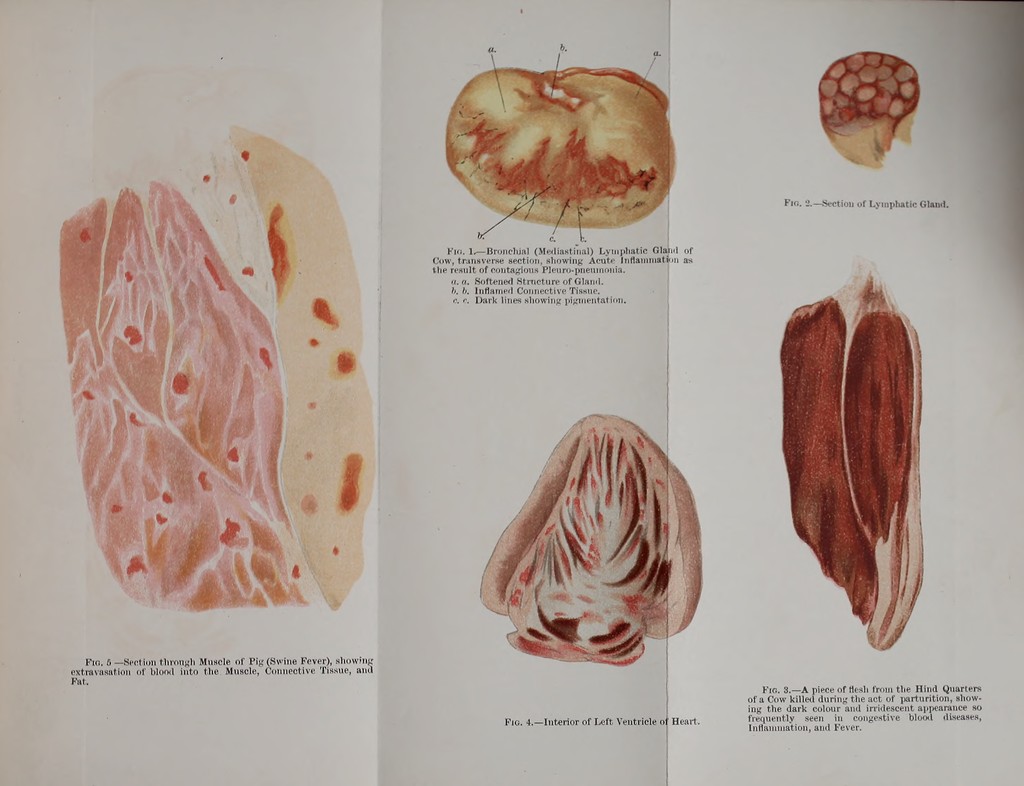
![Fig. 6 — Section through Muscle of Pig (Swine Fever), showing extravasation of blood into the Muscle, Connective Tissue, ami Kill. Fio. 2.—Section of Lymphatic Gland. Fio. 1>—Bronchia] (Mediastinal) Lymphatic Glaii.l of Cow, tr.insverse section, showing Acute Inflatnmat>in as the result of contagious Pleuro-piieumonia. a. a. Softened Structure of Glaml. h. luflatneil Connective Tissue. C r. Dark lines showing pigmentation. ) Fig. 4.—Interior of Left Ventricle of Heart. Fio. 3.—A piece of tiesh from tlie Hind Quarlers of a Cow killed during the act of parturition, show- ing the dark colour and irridescent appearance so frequently seen in congestive blood diseases, Inflammation, and Fever.](https://iiif.wellcomecollection.org/image/b21962820_0039.jp2/full/800%2C/0/default.jpg)