The menstruation and ovulation of Macacus rhesus : with observations on the changes undergone by the discharged follicle. Pt. II / by Walter Heape ; [communicated by M. Foster].
- Heape, Walter, 1855-1929.
- Date:
- 1897
Licence: Public Domain Mark
Credit: The menstruation and ovulation of Macacus rhesus : with observations on the changes undergone by the discharged follicle. Pt. II / by Walter Heape ; [communicated by M. Foster]. Source: Wellcome Collection.
Provider: This material has been provided by The Royal College of Surgeons of England. The original may be consulted at The Royal College of Surgeons of England.
36/40 (page 166)
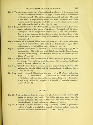
![Fig-. 12. A piece of the follicle represented in fig. 7, showing the minute branched nuclei embedded in a matrix, which fills the cavity of the follicle, the branched ceils of the follicle wall are also shown. (Zeiss, 0, occ. 4.) Fig, 13. A piece of the follicle represented in fig. 8 ; the tissue in the cavity of the follicle is a very loose, wide-meshed connective tissue, continuous with the branched cells of the follicle wall. (Zeiss, 0, occ. 4.) Fig. 14. A piece of the follicle wall represented in fig. 9. The material in the cavity of this follicle is not cellular, but consists of a network of finely granulated material. A few red blood corpuscles lie within its meshes near the wall of the follicle, but there is no blood clot. The wall of the follicle is formed of branching cells, except along its internal edge, and here it is composed of longitudinal, finely drawn out cells, sharply marked off from the follicular cavity, but continuous with the branched processes of the deeper seated cells of the wall. (Zeiss, C, occ. 4.) Fig. ] 5. A piece of the apex of the follicle drawn in fig. 9, together with the tissue intervening between it and the surface of the ovary. The ovarian epithelium is absent at the point x, and is much flattened on either side of this point. Between the apex of the follicle and the point x the tissue is loose and open; it consists of a wide-meshed connective tissue with few nuclei, and many extravasated red-blood corpuscles are scattered about therein, (Zeiss, A, occ. 4.) Fig. 16. A piece of the follicle represented in fig. 10, showing the branched nuclei of the tissue contained within the follicle, and the early formation, in the follicle wall, of nests of nuclei surrounded by branched connective tissue cells. (Zeiss, C, occ. 4.)](https://iiif.wellcomecollection.org/image/b22392701_0038.jp2/full/800%2C/0/default.jpg)