A text-book of pathological histology : an introduction to the study of pathological anatomy / By Dr. Edward Rindfleisch ... Translated from the 2d German ed. ... by William C. Kloman, M.D., assisted by F.T. Miles.
- Rindfleisch, Georg Eduard von, 1836-1908. Lehrbuch der pathologischen gewebelehre zur einfuhrung in das studium der pathologischen anatomie. English
- Date:
- 1872
Licence: Public Domain Mark
Credit: A text-book of pathological histology : an introduction to the study of pathological anatomy / By Dr. Edward Rindfleisch ... Translated from the 2d German ed. ... by William C. Kloman, M.D., assisted by F.T. Miles. Source: Wellcome Collection.
Provider: This material has been provided by the University of Massachusetts Medical School, Lamar Soutter Library, through the Medical Heritage Library. The original may be consulted at the Lamar Soutter Library at the University of Massachusetts Medical School.
106/708 (page 100)
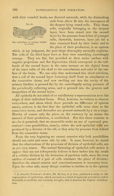
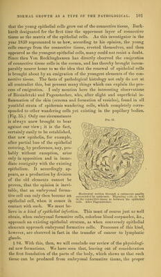
![with their rounded heads, are directed outwards, while the diminishing ends from above fit into the interspaces of FlG-34 the deepest lying round cells. That these cells, originally belonging to the deepest layer, have been raised into the second layer by the pressure from below of younger cells, therewith, however, have yet for a The epithelium tf.urinary bladder tjme remajned fixed by thejr ]ower end to the place of their production, is an opinion which, in my judgment, the pear-shape thoroughly naturally explains. The cells of the third layer have at first view something exceedingly strange. They are flat, but provided at their under surface with angular projections and flat depressions, which correspond to the cell- heads of the second layer, in the same manner as the digital fossae of the inner table of the skull to the convolutions and sulci of the sur- face of the brain. We can only thus understand this third cell-form, that a cell of the second layer loosening itself from its attachment to the connective tissue, and now swelling out into the cavity of the urinary bladder, is pressed flat by the centrifugally acting pressure of the periodically collecting urine, and is pressed into the grooves and inequalities of the second layer. All epithelia do not admit of so satisfactory a representation as to the origin of their individual forms. What, however, we believe to observe everywhere, and about which there prevails no difference of opinion among authors, is the fact that the epithelial cells arise close to the connective tissue, and thereafter are pressed outwards by a vis a tergo. Hereby of course only the place, on the contrary by no means the manner of their production, is established. For this there remains to us—be it premised, that we meanwhile make no use of equivocal gen- eration*—two possibilities, namely, either the new epithelial cells are ^produced by a division of the old, or they arise by pressure from behind -;from the connective tissue. From the very beginning we cannot conceive why both possibilities ^ should not exist and occur side by side. It must, however, be stated that the observations of the processes of division of epithelial cells, are as yet very scarce. The mutual flattening of epithelial cells makes it appear that one not infrequently believes to have before him the result of a cellular division by the formation of partition walls, since the even surface of contact of a pair of cells simulates the plane of division ; therefore the utmost caution and conscientiousness is necessary here. Upon the other side, many things combine to incline us to the opinion * J. Arnold (Virchow's Archiv, Bel. 46) has, it Is true, published a study on the regeneration of epithelium, which amounts to a kind of equivocal generation, and is therewith so carefully and concisely wrought, that it deserves the highest considera- tion.](https://iiif.wellcomecollection.org/image/b21198019_0106.jp2/full/800%2C/0/default.jpg)