On the corpuscles of the blood / by Martin Barry. Pts. [I]-III.
- Martin Barry
- Date:
- 1840-1841
Licence: Public Domain Mark
Credit: On the corpuscles of the blood / by Martin Barry. Pts. [I]-III. Source: Wellcome Collection.
Provider: This material has been provided by The Royal College of Surgeons of England. The original may be consulted at The Royal College of Surgeons of England.
11/112 page 603
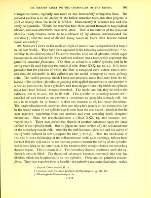
![transparent centre, regularly and more or less transversely arranged in lines. The pellucid nucleus is in the interior of the hollow muscular fibre, and often projects in part or wholly when the latter is divided. Subsequently it becomes less and less easily recognizable. Within the muscular fibre there present themselves longitudinal fibrillse, and soon afterwards transverse striae. That in the interior of the muscular fibre the cavity remains, seems to be confirmed by my already communicated ob- servation'!', that the ends of divided living muscular fibres often become turned inside outwards.^” 30. Schwann’s views on the mode of origin of muscle have been published at length in his late work§. They have since appeared in the following condensed form : “ Ac- cording to the observations of Valentin, muscles arise out of globules, which apply themselves to one another in rows and then coalesce into a fibre, which represents the primitive muscular fasciculus. The fibre so arisen is a hollow cylinder, and in its cavity there lie near together the nuclei of cells (Plate XXX. fig. 18. a.). It is hence probable that the globules of which the fibre is composed were hollow, that is cells, and that the cell-nuclei in this cylinder are the nuclei belonging to those primary cells. The earlier process, which I have not observed, must thus have been the fol- lowing : The (hollow) globules or primary cells applied themselves to one another in a row, or coalesced to form a cylinder; and then the partitions—by which the cylinder must have been divided—became absorbed. The nuclei are flat, they lie within the cylinder, not in its axis, but in its wall. This cylinder or secondary muscle-cell— rounded off and closed at one extremity—continues to grow like a simple cell; but only in its length, for in breadth it does not increase at all, but rather diminishes. The longitudinal growth, however, does not take place merely at the extremities, but in the whole extent of the cylinder; as is seen from the cell-nuclei—which at first lie near together—separating from one another, and even becoming much elongated themselves. Thus the muscle-fasciculus a (Plate XXX. fig. 18.) becomes con- verted into (3. There now occurs the deposit of another substance upon the inner surface of the cylinder-wall,—that is [upon the inner surface of] the cell-membrane of the secondary muscle-cell,—whereby the wall becomes thickened and the cavity of the cylinder reduced in size (compare the fibre y with (3). That the thickening of the wall is not a thickening of the cell-membrane itself, as in cartilage, follows from the fact that the cell-nuclei do not become pushed towards the cavity of the cylinder, but remain lying at the outer part, in the situation they occupied before the secondary deposit began. This is shown in c5. The secondary deposit continues until the cy- linder is entirely filled. The deposited substance becomes converted into very fine fibrillse, which run longitudinally in the cylinder. These are the primitive muscle- jibres. They thus together form a bundle—the primitive muscular fasciculus—which f Hecker’s Neue Annalen, II. 71. J Valentin, in R. Wagner’s Lehrbuch der Physiologie, I. pp. 137.138. § Mikroskopische Untersuchungen, &c. 4 H 2](https://iiif.wellcomecollection.org/image/b22296785_0013.jp2/full/800%2C/0/default.jpg)


 No text description is available for this image
 No text description is available for this image
 No text description is available for this image