Licence: Public Domain Mark
Credit: Further observations on Pareiasaurus / by H.G. Seeley. Source: Wellcome Collection.
Provider: This material has been provided by The Royal College of Surgeons of England. The original may be consulted at The Royal College of Surgeons of England.
8/76 (page 316)
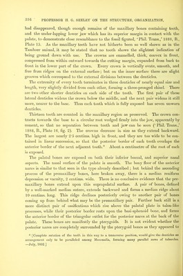
![had disappeared, though enough remains of tlie maxillary bones containing teeth, and the under-lapping lower jaw which has its superior margin in contact with the palate, to demonstrate close resemblance to the fossil figured, ‘Phil. Trans.,’ 1888, B., Plate 15. As the maxillary teeth have not hitherto been so well shown as in the Tamboer animal, it may be stated that no tooth shows the slightest indication of being ground down with use. The crowns are enamelled, thick, convex in front, compressed from within outward towards the cutting margin, expanded from back to front in the lower part of the crown. Every crown is verticallj’’ ovate, smooth, and free from ridges on the external surface; but on the inner surface there are slight grooves which correspond to the external divisions between the denticles. The extremity of every tooth terminates in three denticles of nearly equal size and length, very slightly divided from each other, forming a three-pronged chisel. There are two other shorter denticles on each side of the tooth. The first pair of these lateral denticles widens the crown below tlie middle, and the next pair widens it still more, nearer to the base. Thus each tooth which is fully exposed has seven unworn denticles. Thirteen teeth are counted in the maxillary region as preserved. The crown con- tracts towards the base to a circular root wedged firmly into the jaw, apparently by cement, so that no separation between tooth and jaw can be seen (‘ Phil. Trans.,’ 1888, B., Plate 16. fig. 2). The crowns decrease in size as they extend backward. The largest are nearly 2'5 centims. high in front, and they are too wide to be con- tained in linear succession, so that the posterior border of each tooth overlaps the anterior border of the next adjacent tooth.* About a centimetre of the root of each is exposed. The palatal bones are exj^osed on both their inferior buccal, and superior nasal aspects. The nasal surface of the palate is smooth. The bony floor of the anterior nares is similar to that seen in the type already described ; but behind the ascending process of the premaxillary bones, here broken away, there is a median reniform depression or vacuity, 2 centims. wide. There is no conclusive evidence that the pre- maxillary bones extend upon this suprapalatal surface, A pair of bones, defined by a well-marked median suture, extends backward and forms a median ridge about 10 centims. long. This ridge declines posteriorly owing to another pair of bones coming up from behind what may be the premaxillary pair. Further back still is a more distinct pair of ossifications which rise above the palatal plate in talon-like processes, while their posterior border rests upon the basi-sphenoid bone, and forms the anterior border of the triangular outlet for the posterior nares at the back of the palate. These bones are manifestly the pterygoids. It is not evident whether the posterior nares are completely surrounded hy the pterygoid bones as they appeared to * [Complete rotation of the teetti in this way to a transverse position, would give the denticles an arrangement only to be jiaralleled among Mammalia, forming many parallel rows of tubercles. —July, 1892.]](https://iiif.wellcomecollection.org/image/b22417278_0010.jp2/full/800%2C/0/default.jpg)