Some account of the last yellow fever epidemic of British Guiana / by Daniel Blair ; edited by John Davy.
- Date:
- 1850
Licence: Public Domain Mark
Credit: Some account of the last yellow fever epidemic of British Guiana / by Daniel Blair ; edited by John Davy. Source: Wellcome Collection.
Provider: This material has been provided by the Royal College of Physicians of Edinburgh. The original may be consulted at the Royal College of Physicians of Edinburgh.
120/222 (page 100)
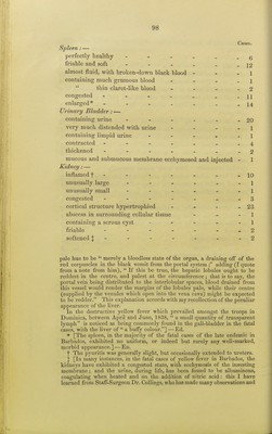
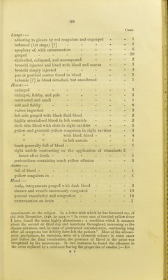
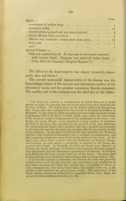
![, Cases. Head:— membranes of yellow tinge - - - . j arachnoid milky - - . . . - 11 choroid plexus gorged and pia mater injected . 2 serous effusion from arachnoid - - - - 3 effusion into ventricles (seldom more than giss) - - 13 brain pale ] soft *- - ... Spinal Column:— Only one examination of. In that case it was found congested with venous blood. Priapism was observed before death (Case 2816 of Seaman's Hospital Register). I The Blood in the dead subjects was almost invariably abnor- mally thin and black, f The second anatomical characteristic of the disease was the hemorrhagic lesion of the mucous and submucous surface of the alimentary canal, and the peculiar excretions thei'ein contained. The cardiac end of the stomach was the chief site of the inflam- * [As there are varieties or modifications of yellow fever, so it would appear, as might be expected, that the lesions discovered after death are also far from uniform. The details given by the Author sufficiently demonstrate this. I may add a few examples from the records in the Inspector's Office, in Barbados. In the endemic which prevailed, and was so fatal among the troops at Brimstone Hill in St. Kitt's, in 1812, which came under the observation of Dr. Jackson, marks of inflammation of the membranes of the brain — viz., lymph and serum effused—were noticed in a large number of instances. In 1811, in the fever that prevailed in the garrison of St. Ann, Barbados, Dr. (afterwards Sir Charles) Ker mentions that there was little determination to the stomach, but that the brain was strongly affected, the patient usually dying comatose. In the fever of 1816, in the same garrison, Dr. Ferguson reports — that cerebral inflammation was a common accompaniment., and that coagulable lymph was often found on the surface of the brain. In the same year yellow fever occurred and was destructive in Antigua, Tobago, and Grenada: at each station a peculiarity is reported in the lesions dis- covered after death. In Grenada, 1 he liver was found most affected: in Tobago, the throat and lungs; in the latter, the intestines often had a gangre- nous appearance, and the omentum an appearance a's if inflamed : in Antigua, as in Barbados, the brain was the chief seat of the lesion. The worst cases in this epidemic were marked by haemorrhages and the black vomit.]—En. f [In the late endemic in Barbados, the blood in the body was generally of dark colour, as if not duly aerated ; but, according to my recollection, it was rather unctuous and thick than unduly thin : concretions of fibrin, or even crassamentum, were rarely found where they usually occur, viz., in the cavities of the heart and the great venous trunks. The appearance of the blood, whether abstracted during life or examined shortly after death, did not at all denote inflammatory action.]—Ed.](https://iiif.wellcomecollection.org/image/b21976077_0120.jp2/full/800%2C/0/default.jpg)